Comparative Study
. 2004 Oct 15;18(20):2545-56.
doi: 10.1101/gad.1245904.

The geminivirus nuclear shuttle protein is a virulence factor that suppresses transmembrane receptor kinase activity

Affiliations
Comparative Study

The geminivirus nuclear shuttle protein is a virulence factor that suppresses transmembrane receptor kinase activity

Elizabeth P B Fontes et al. Genes Dev. .

Abstract

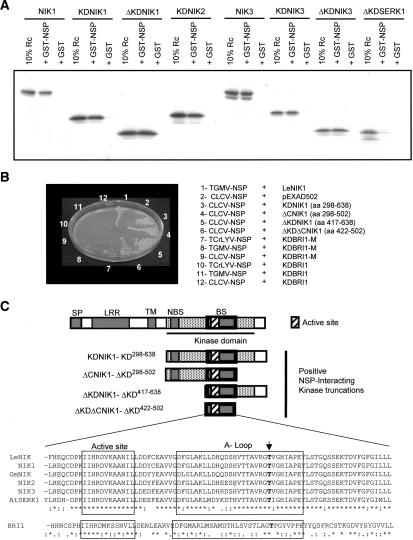
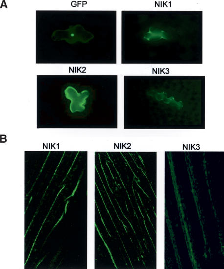
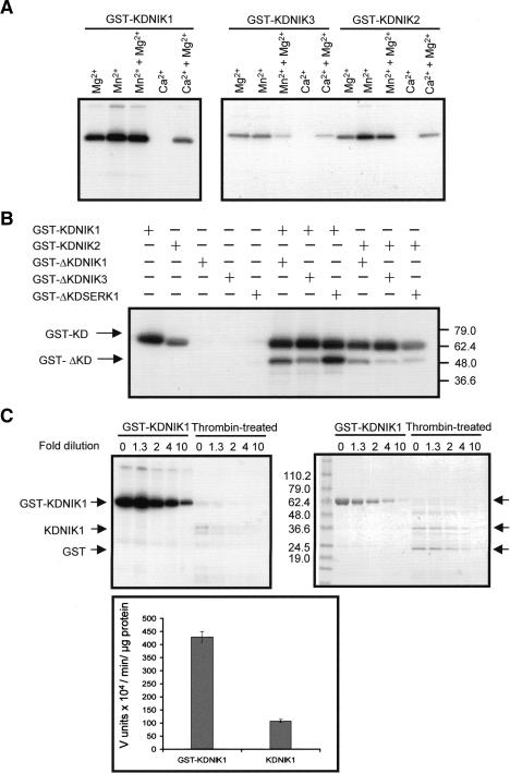
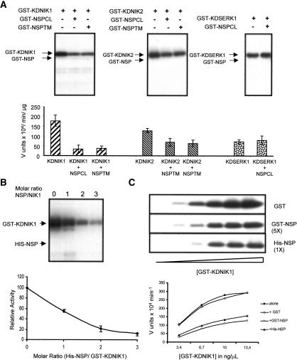
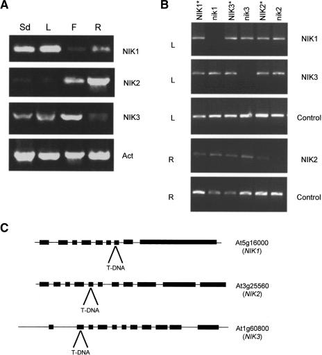
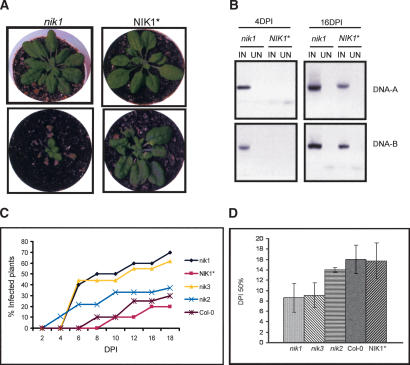
Despite the large number of leucine-rich-repeat (LRR) receptor-like-kinases (RLKs) in plants and their conceptual relevance in signaling events, functional information is restricted to a few family members. Here we describe the characterization of new LRR-RLK family members as virulence targets of the geminivirus nuclear shuttle protein (NSP). NSP interacts specifically with three LRR-RLKs, NIK1, NIK2, and NIK3, through an 80-amino acid region that encompasses the kinase active site and A-loop. We demonstrate that these NSP-interacting kinases (NIKs) are membrane-localized proteins with biochemical properties of signaling receptors. They behave as authentic kinase proteins that undergo autophosphorylation and can also phosphorylate exogenous substrates. Autophosphorylation occurs via an intermolecular event and oligomerization precedes the activation of the kinase. Binding of NSP to NIK inhibits its kinase activity in vitro, suggesting that NIK is involved in antiviral defense response. In support of this, infectivity assays showed a positive correlation between infection rate and loss of NIK1 and NIK3 function. Our data are consistent with a model in which NSP acts as a virulence factor to suppress NIK-mediated antiviral responses.


Figures

Figure 1.
Figure 1.
Specificity of NSP interaction with LRR-RLKs. (A) In vitro interaction of NSP with NIKs. In vitro transcribed and translated 35S-labeled proteins, as indicated in the figure, were allowed to interact with bacterially expressed GST or GST-NSP linked to glutathione-Sepharose beads. After extensive washing of the beads, the retained proteins were separated by SDS-PAGE and visualized through fluorography. Input contains a sample (10% reaction) of the respective transcription and translation reaction mixtures. (B) NSP does not interact with either active or inactive kinase domain of BRI1, a member of the LRR IX subfamily of the RLK family. Yeast cells expressing the indicated recombinant proteins were plated on selective medium lacking leucine, tryptophan, uracil, and histidine and supplemented with 25 mM 3-AT and grown for 3 d at 30°C. (C) Mapping of NSP-binding domain on NIK1. The diagram represents the NIK1 truncated versions that interacted with NSP (B). In the sequence comparison of the NSP-binding domain among NIKs and AtSERK1- and BRI1-corresponding regions, the active site and A-loop are boxed and the arrow indicates the conserved threonine residue. (SP) Signal peptide; (LRR) leucine-rich repeats; (TM) transmembrane domain; (NBS) nucleotide-binding site; (BS) NSP-binding site.
Figure 2.
Figure 2.
NIK1, NIK2, and NIK3 are localized to the plasma membrane. (A) Fluorescence deconvolution microscopy images from epidermal cells of Arabidopsis leaves bombarded with GFP, NIK1–GFP, NIK2–GFP, and NIK3–GFP, under the control of the 35S promoter. (B) Confocal fluorescence images of Arabidopsis cauline cells stably transformed with NIK1–GFP, NIK2–GFP, or NIK3–GFP.
Figure 3.
Figure 3.
The NIKs exhibit biochemical properties of signaling receptors. (A) NIKs undergo autophosphorylation in vitro in a Mg2+- or Mn2+-dependent reaction. Bacterially produced GST-fusion proteins (as indicated) were purified and aliquots of 200–500 ng were incubated with [γ-32P]ATP in the presence of the indicated cations. After separation on 4%–20% SDS-PAGE, the phosphoproteins were visualized by autoradiography. (B) Autophosphorylation of NIKs occurs intermolecularly. GST-fusion proteins (as indicated) were incubated with [γ-32P]ATP, separated by SDS-PAGE, and visualized by autoradiography. The migrations of GST-fused kinase domains (GST-KD) and their truncated versions (GST-ΔKD) are indicated. The positions of molecular markers are indicated on the right in kilodaltons. (C) Oligomerization is required for kinase activation. GST-KDNIK1 was cleaved with thrombin and equal amounts of intact or thrombin-cleaved GST-KDNIK1 were used as serial dilutions in the kinase assay. Phosphorylated proteins were analyzed by Coomassie-stained SDS-PAGE (right) and visualized by autoradiography (left). The migrations of GST-KDNIK and thrombin-cleaved GST-KDNIK1 (GST and KDNIK1) are indicated. Molecular markers are shown in kilodaltons. The plot on the bottom shows the relative kinase activity of GST-KDNIK1 and thrombin-cleaved GST-KDNIK1 expressed as specific activity.
Figure 4.
Figure 4.
NSP inhibits the kinase activity of NIKs. (A) Both TGMV NSP and CaLCuV NSP inhibit NIK1 and NIK2 kinase activity in vitro. GST-KDNIK1 or GST-KDNIK2 were incubated with [γ-32P]ATP and GST-NSP from CaLCuV (GST-NSPCL) or from TGMV (GST-NSPTM). GST-KDSERK1 was incubated with GST-NSPCL. After separation on SDS-PAGE, phosphoproteins were visualized by autoradiography and quantified by phosphoimaging. Relative values of 32P incorporation are the mean of three replicas. (B) Stoi-chiometry of inhibition. Purified GST-KDNIK1 (250 ng) was incubated with [γ-32P]ATP in the presence of increasing amounts of His-NSP. The plot of protein phosphorylation versus molar excess of His-GST was from three replicas. (C) Kinetics of inhibition. Increasing amounts of GST-KDNIK1 were incubated with [γ-32P]ATP in the presence of GST (5 ng/μL), His-NSP (5 ng/μL), or GST-NSP (40 ng/μL). NIK1 phosphorylation was quantified and plotted versus NSP protein concentration.
Figure 5.
Figure 5.
NIKs expression pattern and knockout lines. (A) Organ-specific expression of NIKs. RT–PCR was performed with cDNA prepared from seedlings (Sd), leaves (L), flowers (F), and roots (R) RNA with gene-specific primers, as indicated on the right. (B) Knockout lines for NSP-interacting transmembrane receptors. RT–PCR was performed on leaf (L) and root (R) RNA samples from wild-type (NIK1, NIK2, NIK3), nik1, nik2, and nik3 plants with gene-specific primers, as indicated on the right. The asterisks indicate that the homozygous wild-type alleles were recovered in segregating lines of T-DNA insertion mutants. (C) Annotated NIK genomic loci and diagram of T-DNA insertions. The genes are indicated in the 5′–3′ orientation. Black boxes represent the exons. The position of T-DNA insertion in the null alleles is indicated.
Figure 6.
Figure 6.
NIK knockout lines exhibit enhanced susceptibility to geminivirus infection. (A) Symptoms associated with CaLCuV infection in knockout lines. Tandemly repeated viral DNA-A and DNA-B were introduced into plants by biolistic inoculation. NIK1 (bottom right) and nik1 (bottom left) infected with CaLCuV at 18 DPI. On the top, NIK1 (right) and nik1 (left) are mock-inoculated plants. (B) Viral DNA accumulation in infected lines. Total DNA was isolated from greenhouse grown NIK1 and nik1 plants at 4 and 16 DPI and subjected to DNA blot analysis with 32P-labeled DNA-A or DNA-B probes, as indicated on the right. (IN) Viral DNA-inoculated plants; (UN) mock-inoculated plants. (C) The onset of infection is accelerated in nik1 and nik3 knockout lines. Ecotype Col-0, nik1, NIK1, nik2, and nik3 lines were infected with CaLCuV DNA by the biolistic method. Values represent the percent of systemically infected plants at different DPI. (D) Infection rates in nik null alleles. The infection rate was expressed as DPI to get 50% of infected plants. Values for DPI50% are the mean ± standard deviation from three replicas.

Similar articles

Cited by

References

    1. Alonso J.M., Stepanova, A.N., Leisse, T.J., Kim, C.J., Chen, H., Shinn, P., Stevenson, D.K., Zimmerman, J., Barajas, P., Cheuk, R., et al. 2003. Genome-wide insertional mutagenesis of Arabidopsis thaliana. Science 301: 653-657. - PubMed
    1. The Arabidopsis Genome Initiative. 2000. Analysis of the genome sequence of the flowering plant Arabidopsis thaliana. Nature 408: 796-815. - PubMed
    1. Cascardo J.C.M., Almeida, R.S., Buzeli, R.A.A., Carolino, S.M.B., Otoni, W.C., and Fontes, E.P.B. 2000. The phosphorylation state and expression of soybean BiP isoforms are differentially regulated following abiotic stresses. J. Biol. Chem. 275: 14494-14500. - PubMed
    1. Clark S.E., Williams, R.W., and Meyerowitz, E.M. 1997. The CLAVATA1 gene encodes a putative receptor kinase that controls shoot and floral meristem size in Arabidopsis. Cell 89: 575-585. - PubMed
    1. Dievart A. and Clark, S.E. 2004. LRR-containing receptors regulating plant development and defense. Development 131: 251-261. - PubMed

Publication types

MeSH terms