. 2004 Oct;14(10B):2111-20.
doi: 10.1101/gr.2512204.

Concerted assembly and cloning of multiple DNA segments using in vitro site-specific recombination: functional analysis of multi-segment expression clones

Affiliations

Concerted assembly and cloning of multiple DNA segments using in vitro site-specific recombination: functional analysis of multi-segment expression clones

David L Cheo et al. Genome Res. 2004 Oct.

Abstract

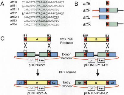
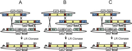
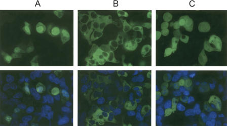
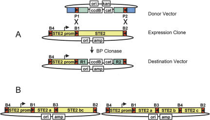
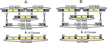
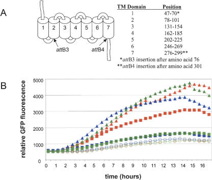
The ability to clone and manipulate DNA segments is central to molecular methods that enable expression, screening, and functional characterization of genes, proteins, and regulatory elements. We previously described the development of a novel technology that utilizes in vitro site-specific recombination to provide a robust and flexible platform for high-throughput cloning and transfer of DNA segments. By using an expanded repertoire of recombination sites with unique specificities, we have extended the technology to enable the high-efficiency in vitro assembly and concerted cloning of multiple DNA segments into a vector backbone in a predefined order, orientation, and reading frame. The efficiency and flexibility of this approach enables collections of functional elements to be generated and mixed in a combinatorial fashion for the parallel assembly of numerous multi-segment constructs. The assembled constructs can be further manipulated by directing exchange of defined segments with alternate DNA segments. In this report, we demonstrate feasibility of the technology and application to the generation of fusion proteins, the linkage of promoters to genes, and the assembly of multiple protein domains. The technology has broad implications for cell and protein engineering, the expression of multidomain proteins, and gene function analysis.


Figures

Figure 1
Figure 1
Summary of att sites and BP cloning. (A) List of attB sites; attB0 is the E. coli λ bacteriophage attachment site (Landy 1989). Shaded area indicates 7-bp overlap (Int cut sites); underlined bases indicate changes from attB0. (B) Schematic diagram of recombination sites. (Red rectangles) attB sites, the orientation of the asymmetric overlap region is indicated by the triangle. (Blue and green rectangles) attL and attR arms, respectively. (Red line) vector backbone segment; (black line) ccdB counter selection gene (Bernard and Couturier 1992) and chloramphenicol acetyltransferase (cat) selection marker. (C) Examples of BP cloning of attB PCR products into Donor Vectors to generate Entry Clones. Note that reversing the orientations of both the attB1 site in the PCR product and attP1 site in the Donor Vector results in an attR1 site in the Entry Clone.
Figure 2
Figure 2
Overview of two-segment cloning. (A) Linkage of a 3′ element Entry Clone (R2-L3) to standard Entry Clones (L1-L2) and cloning into a Destination Vector (R1-R3). (B) Linkage of a 5′ element Entry Clone (L4-R1) to standard Entry Clone and cloning into a Destination Vector (R4-R2). (C) Alternate two-segment cloning strategy linking two Entry Clones (L1-L3 and R3-L2) and cloning into a standard Destination Vector (R1-R2).
Figure 3
Figure 3
Fluorescent, confocal, live-cell imaging of HEK-293 cells transfected with (A) NLS-eGFP, (B) β-Arrestin-eGFP fusions, or (C) untagged eGFP. Forty-eight hours post-transfection, cells were stained with Hoechst 33342 and imaged confocally at 60× magnification on a Pathway HT system (Atto Bioscience). Pseudo-colored images of a typical Z-stack section are shown. (Top) GFP channel alone; (bottom) Hoechst + GFP.
Figure 4
Figure 4
Functional analysis of transcriptional fusions. (A) Promoter-eGFP fusions and (B) promoter-STE2 receptor fusions in yeast cells co-transformed with the FUS1-yeGFP reporter. Relative GFP fluorescence was measured over a 16-h period. Yeast strains were assayed in the presence (filled symbols) or absence (open symbols) of 500 nM α-factor as described in the Methods. Promoter fusions are as follows: STE2 (Red), ADH1 (yellow), GPD1 (blue), TEF2 (green), and CUP1 (black). Yeast strains containing the CUP1 promoter fusions were assayed in the presence (black circles) and absence (black squares) of 100 μM copper sulfate.
Figure 6
Figure 6
STE2 Expression Constructs (A) Reaction of the B1-B2 sites in a multi-segment Expression Clone with a Donor Vector (P1-P2) to generate a STE2 promoter Destination Vector. (B) Diagram of STE2 receptor constructs resulting from 2 and 3 segment cloning into the Destination Vector shown in (A).
Figure 5
Figure 5
Overview of three-segment cloning. (A) Linkage of a 5′ Entry Clone, a standard (L1-L2) Entry Clone, and a 3′ Entry Clone and a Destination Vector (R4-R3). (B) Alternate three-segment cloning strategy using a standard Gateway Destination Vector (R1-R2).
Figure 7
Figure 7
Functional analysis of STE2 receptor constructs (A) Ste2 transmembrane topology (Parrish et al. 2002) and att site locations. (B) Response to α-factor treatment of yeast strains containing STE2 receptor constructs cotransformed with the FUS1-yeGFP reporter. The STE2 receptor constructs were expressed using the STE2 promoter. Relative GFP fluorescence was measured over a 16-h period. Yeast strains were assayed in the presence of 500 nM (triangles), 50 nM (squares), or absence (circles) of α-factor as described in the Methods. Symbols are as follows: wild-type Ste2 receptor (red), Ste2 receptor containing att3 (blue), and Ste2 receptor containing both attB3 and attB4 (green).

References

    1. Barak, L.S., Ferguson, S.S., Zhang, J., and Caron, M.G. 1997. A β-arrestin/green fluorescent protein biosensor for detecting G protein-coupled receptor activation. J. Biol. Chem. 272: 27497-27500. - PubMed
    1. Bardwell, L., Cook, J.G., Inouye, C.J., and Thorner, J. 1994. Signal propagation and regulation in the mating pheromone response pathway of the yeast Saccharomyces cerevisiae. Dev. Biol. 166: 363-379. - PubMed
    1. Baudin, A., Ozier-Kalogeropoulos, O., Denouel, A., Lacroute, F., and Cullin, C. 1993. A simple and efficient method for direct gene deletion in Saccharomyces cerevisiae. Nucleic Acids Res. 21: 3329-3330. - PMC - PubMed
    1. Bernard, P. and Couturier, M. 1992. Cell killing by the F plasmid CcdB protein involves poisoning of DNA-topoisomerase II complexes. J. Mol. Biol. 226: 735-745. - PubMed
    1. Boulton, S.J., Gartner, A., Reboul, J., Vaglio, P., Dyson, N., Hill, D.E., and Vidal, M. 2002. Combined functional genomic maps of the C. elegans DNA damage response. Science 295: 127-131. - PubMed

WEB SITE REFERENCES

    1. http://www.fruitfly.org/EST/gateway.shtml; Drosophila ORFs.
    1. http://xgc.nci.nih.gov; Xenopus ORFs.
    1. http://zgc.nci.nih.gov; zebrafish ORFs.
    1. http://mgc.nci.nih.gov; human, mouse and rat ORFs.

Publication types

LinkOut - more resources