

Molecular methods for diagnosis of viral encephalitis
- PMID: 15489354
- PMCID: PMC523566
- DOI: 10.1128/CMR.17.4.903-925.2004
Molecular methods for diagnosis of viral encephalitis
Abstract
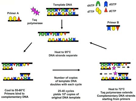
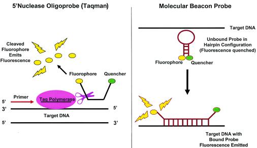
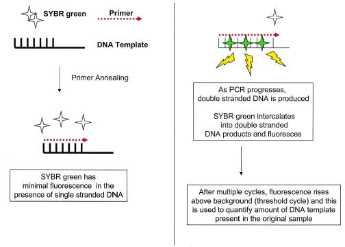
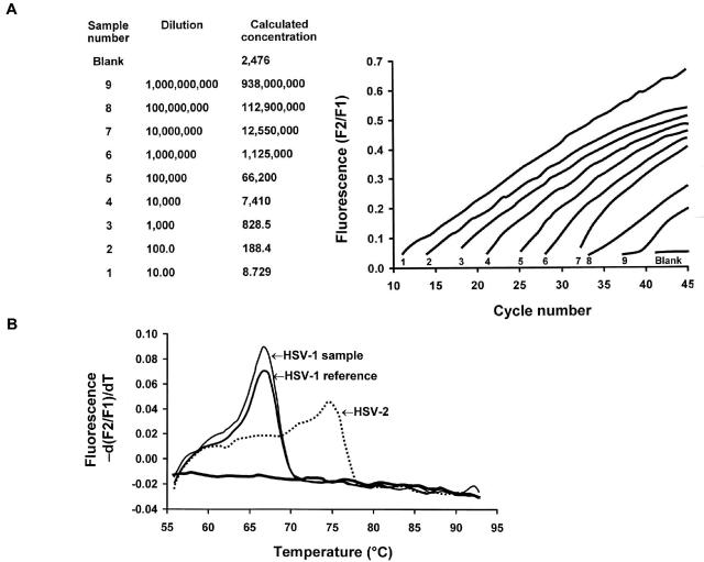
Hundreds of viruses cause central nervous system (CNS) disease, including meningoencephalitis and postinfectious encephalomyelitis, in humans. The cerebrospinal fluid (CSF) is abnormal in >90% of cases; however, routine CSF studies only rarely lead to identification of a specific etiologic agent. Diagnosis of viral infections of the CNS has been revolutionized by the advent of new molecular diagnostic technologies to amplify viral nucleic acid from CSF, including PCR, nucleic acid sequence-based amplification, and branched-DNA assay. PCR is ideally suited for identifying fastidious organisms that may be difficult or impossible to culture and has been widely applied for detection of both DNA and RNA viruses in CSF. The technique can be performed rapidly and inexpensively and has become an integral component of diagnostic medical practice in the United States and other developed countries. In addition to its use for identification of etiologic agents of CNS disease in the clinical setting, PCR has also been used to quantitate viral load and monitor duration and adequacy of antiviral drug therapy. PCR has also been applied in the research setting to help discriminate active versus postinfectious immune-mediate disease, identify determinants of drug resistance, and investigate the etiology of neurologic disease of uncertain cause. This review discusses general principles of PCR and reverse transcription-PCR, including qualitative, quantitative, and multiplex techniques, with comment on issues of sensitivity, specificity, and positive and negative predictive values. The application of molecular diagnostic methods for diagnosis of specific infectious entities is reviewed in detail, including viruses for which PCR is of proven efficacy and is widely available, viruses for which PCR is less widely available or for which PCR has unproven sensitivity and specificity, and nonviral entities which can mimic viral CNS disease.
Figures
References
-
- Aberle, S. W., and E. Puchhammer-Stockl. 2002. Diagnosis of herpesvirus infections of the central nervous system. J. Clin. Virol. 25(Suppl. 1):S79-S85. - PubMed
-
- Abravaya, K., J. Huff, R. Marshall, B. Merchant, C. Mullen, G. Schneider, and J. Robinson. 2003. Molecular beacons as diagnostic tools: technology and applications. Clin. Chem. Lab. Med. 41:468-474. - PubMed
-
- Abzug, M. J., G. Cloud, J. Bradley, P. J. Sanchez, J. Romero, D. Powell, M. Lepow, C. Mani, E. V. Capparelli, S. Blount, F. Lakeman, R. J. Whitley, and D. W. Kimberlin. 2003. Double blind placebo-controlled trial of pleconaril in infants with enterovirus meningitis. Pediatr. Infect. Dis.J. 22:335-341. - PubMed
-
- Abzug, M. J., H. L. Keyserling, M. L. Lee, M. J. Levin, and H. A. Rotbart. 1995. Neonatal enterovirus infection: virology, serology, and effects of intravenous immune globulin. Clin. Infect. Dis. 20:1201-1206. - PubMed
Publication types
MeSH terms
LinkOut - more resources
Full Text Sources
Other Literature Sources
Medical
