. 2004 Nov;186(21):7069-83.
doi: 10.1128/JB.186.21.7069-7083.2004.

Lactobacillus plantarum bacteriophage LP65: a new member of the SPO1-like genus of the family Myoviridae

Affiliations

Lactobacillus plantarum bacteriophage LP65: a new member of the SPO1-like genus of the family Myoviridae

Sandra Chibani-Chennoufi et al. J Bacteriol. 2004 Nov.

Abstract

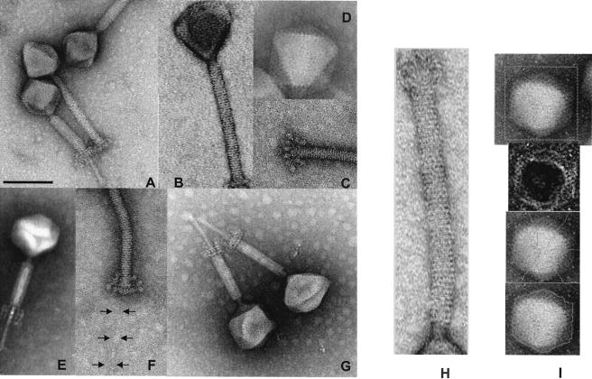
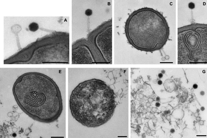
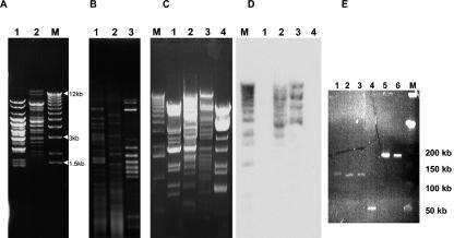
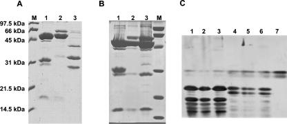
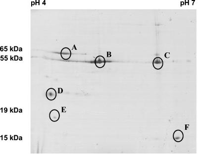
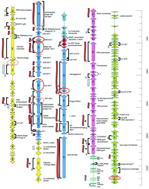
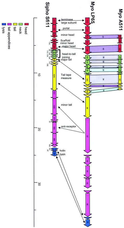
The virulent Lactobacillus plantarum myophage LP65 was isolated from industrial meat fermentation. Tail contraction led to reorganization of the tail sheath and the baseplate; a tail tube was extruded. In ultrathin section the phage adsorbed via its baseplate to the exterior of the cell, while the tail tube tunneled through the thick bacterial cell wall. Convoluted membrane structures were induced in the infected cell. Progeny phage was detected 100 min postinfection, and lysis occurred after extensive digestion of the cell wall. Sequence analysis revealed a genome of 131,573 bp of nonredundant DNA. Four major genome regions and a large tRNA gene cluster were observed. One module corresponded to DNA replication genes. Helicase/primase and two replication/recombination enzymes represented the only links to T4-like Myoviridae from gram-negative bacteria. Another module corresponded to the structural genes. Sequence relatedness identified links with Listeria phage A511, Staphylococcus phage K, and Bacillus phage SPO1. LP65 structural proteins were identified by two-dimensional proteome analysis and mass spectrometry. The putative tail sheath protein showed a shear-induced change in electrophoretic migration behavior. The genome organization of the structural module in LP65 resembled that of Siphoviridae from the lambda supergroup.


Figures

FIG. 1.
FIG. 1.
Negative-staining electron microscopy of CsCl-purified L. plantarum phage LP65. (A) Comparison of LP65 with contracted and noncontracted tails. (B) Striation of the tail sheath. (C) Baseplate of LP65 with noncontracted tail. (D) Icosahedral faces of the phage head. (E) Visualization of a structure within the interior of the tail. (F) Visualization of the tail fiber extending from the baseplate. (G) Transformation of the baseplate after tail contraction. (H) Image analysis of the tail: determination of the average distance of the tail stacks. (I) Image analysis of the head. From top to bottom: original image, processed gradient image, top of the polygon with medians, segmentation of the polyhedron without geometrical constraints. The negative stain was either ammonium molybdate coupled with bacitracin (C, D, F, G, and I), uranyl acetate (A, B, and H), or phosphotungstic acid (E). Bar, 100 nm. For the other dimensions, see Table 2.
FIG. 2.
FIG. 2.
Ultrathin section of LP65-infected L. plantarum cells. (A) LP65 with an empty and a full head adsorbed to the cell wall. Note the small protein shell surrounding the DNA-filled head of the right phage particle and the tail tube of the left particle crossing the cell wall. (B) Phage adsorbing near the septum of a cell, showing the baseplate contacting the surface of the cell wall. (C) Adsorbing LP65 particles. Note again the tail tube and the bulging of the cell wall in the cytoplasm. (D) LP65 making contact via the baseplate with the cell wall and the underlying convolution of cell membranes. (E) Cell containing an intracytoplasmic membrane convolution with phage ghosts adsorbed to the cell wall. (F) A cell that has lost most of its cell wall and is about to release its cytoplasmic contents at the top right. Structures suggestive of phage particles are at the central clearing of the cell. (G) Extruded cytoplasm from a lysed cell with filled heads and empty preheads. Bars, 250 nm.
FIG. 3.
FIG. 3.
DNA analysis of L. plantarum phages. (A) XbaI digestion of DNA from myophage fri (lane 1) and LP65 (lane 2). M: size marker (1-kb lambda DNA ladder; Invitrogen). (B) AccI digests of DNA from phage fri (lane 1), LP65 (lane 2), and the mitomycin C-induced resident prophage LP651 (lane 3) from the propagating L. plantarum strain 65. (C) HindIII digest of DNA from siphophages LP45 (lane 1) and LP76 (lane 4) and myophages fri (lane 2) and LP65 (lane 3). M: size marker (as in A). (D) Southern blot corresponding to the HindIII digest in panel C, probed with radiolabeled phage LP65 DNA. The marker was revealed with labeled lambda DNA. (E) Pulsed-field gel electrophoresis of L. plantarum myophages LP43 (lane 1), LP57 (lane 2), and LP65 (lane 3), beta-4-like coliphage JS122.1 (lane 4), and T4-like coliphages JS148 (lane 5) and RB33 (lane 6). M: size marker (phage lambda concatemers, 50-kb DNA ladder; Promega).
FIG. 4.
FIG. 4.
1-D protein analysis of L. plantarum phages. (A) SDS-PAGE of untreated CsCl-purified myophages LP65 (lane 1) and fri (lane 2) and siphophage LP45 (lane 3). M: broad-range protein marker (Bio-Rad). (B) Effects of ultrasound treatment (lane 2) and syringe shearing (lane 3) of LP65 on the protein pattern in SDS-PAGE gels compared to that for the untreated preparation (lane 1). (C) V8 peptide maps of the 55-kDa proteins of the three LP65 preparations for which results are shown in panel B, lanes 1 to 3 (lanes 1 to 3), the 33-kDa protein from LP65 for which results are shown in panel B, lane 1 (lane 4), and the 65-kDa proteins from LP65 for which results are shown in panel B, lanes 2 and 3 (lanes 5 and 6). The V8 digestion represents untreated (lanes 1 and 4), ultrasound-treated (lanes 2 and 5), or syringe-sheared (lanes 3 and 6) LP65 particles. V8 protease is shown in lane 7.
FIG. 5.
FIG. 5.
2-D protein analysis of phage LP65. Coomassie-stained 2-D gel electrophoresis of CsCl-purified phage LP65. The first dimension used a linear pH 4 to 7 IPG strip gel (Pharmacia), and the second used a 10 to 20% gradient SDS-PAGE gel. Proteins analyzed by nano-ESI-MS (MS/MS) are identified by circles.
FIG. 6.
FIG. 6.
Genome sequence of phage LP65. ORF (arrows) and tRNA (small turquoise bars) prediction in the 131,573-bp sequence of the single contig obtained for LP65 DNA. Gene clusters defined by overall genome organization and database matches are marked with different colors (green, left arm; violet, DNA replication module; red, lysis cassette; blue, structural module; yellow, unknown). Brackets above and below the ORFs indicate matches of the deduced protein with predicted proteins from phages of gram-positive bacteria (except SPO1 and A511) andB. subtilis myophage SPO1, respectively. The matching SPO1 genes are indicated with their gene numbers. Red and grey flags below the ORF indicate significant and weak matches with Staphylococcus phage K, respectively. Genes sharing amino acid sequence identity with T4-like myophages are marked with an asterisk. Gene products identified by MS/MS are circled in red. Dotted circles represent identifications with low scores (Sequest scores of <20).
FIG. 7.
FIG. 7.
Comparative genomics suggests an Sfi11 Siphovirus-like organization of the putative structural gene cluster from LP65. The deduced structural gene map from LP65 (center) is aligned with the corresponding region from Listeria myophage A511 (top). Genes sharing protein sequence identity are linked by different intensities of shading (numbers give the percent amino acid identity). Likewise, the LP65 gene map is also aligned with the structural gene map from S. thermophilus phage Sfi11, the type phage of a widely distributed class of lambda-like Siphoviridae in low-GC-content gram-positive bacteria. The modular structure of the Sfi11 structural gene cluster is indicated by the color code as analyzed previously (31), and selected genes are indicated with their deduced functions. Double-headed arrows link genes possibly sharing the same functions according to database matches obtained for LP65 ORFs. The colinearity of the gene maps from the phages suggests the modular structure for LP65 and A511 indicated by the color code.

Comment in

  • Phage taxonomy: we agree to disagree.
    Nelson D. Nelson D. J Bacteriol. 2004 Nov;186(21):7029-31. doi: 10.1128/JB.186.21.7029-7031.2004. J Bacteriol. 2004. PMID: 15489416 Free PMC article. No abstract available.

References

    1. Ackermann, H. W. 1998. Tailed bacteriophages: the order Caudovirales. Adv. Virus Res. 51:135-201. - PMC - PubMed
    1. Allison, G. E., D. Angeles, N. Tran-Dinh, and N. K. Verma. 2002. Complete genomic sequence of SfV, a serotype-converting temperate bacteriophage of Shigella flexneri. J. Bacteriol. 184:1974-1987. - PMC - PubMed
    1. Altermann, E., J. R. Klein, and B. Henrich. 1999. Primary structure and features of the genome of the Lactobacillus gasseri temperate bacteriophage φadh. Gene 236:333-346. - PubMed
    1. Altschul, S. F., T. L. Madden, A. A. Schaffer, J. Zhang, Z. Zhang, W. Miller, and D. J. Lipman. 1997. Gapped BLAST and PSI-BLAST: a new generation of protein database search programs. Nucleic Acids Res. 25:3389-3402. - PMC - PubMed
    1. Arisaka, F., J. Engel, and H. Klump. 1981. Contraction and dissociation of the bacteriophage T4 tail sheath induced by heat and urea. Prog. Clin. Biol. Res. 64:365-379. - PubMed

Publication types

Associated data

LinkOut - more resources