. 2004 Oct;114(8):1136-45.
doi: 10.1172/JCI21633.

Effect of fetal hemoglobin on microvascular regulation in sickle transgenic-knockout mice

Affiliations

Effect of fetal hemoglobin on microvascular regulation in sickle transgenic-knockout mice

Dhananjay K Kaul et al. J Clin Invest. 2004 Oct.

Abstract

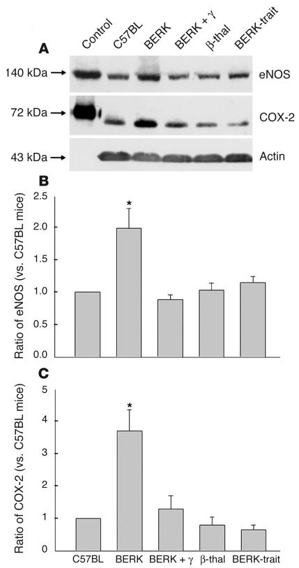
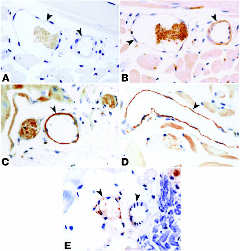
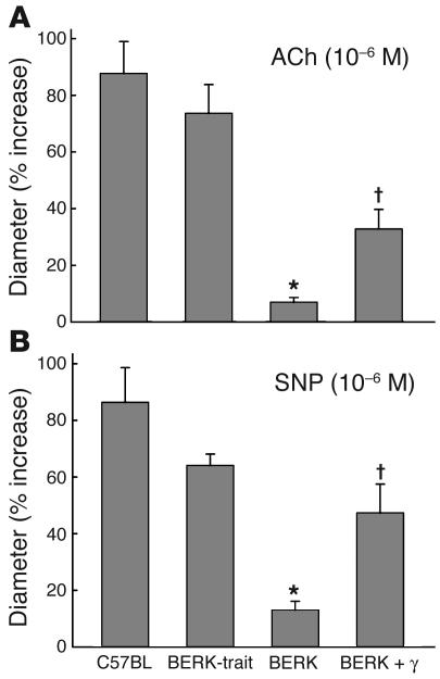
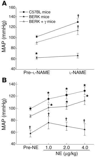
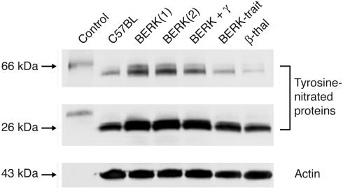
In sickle cell disease, intravascular sickling and attendant flow abnormalities underlie the chronic inflammation and vascular endothelial abnormalities. However, the relationship between sickling and vascular tone is not well understood. We hypothesized that sickling-induced vaso-occlusive events and attendant oxidative stress will affect microvascular regulatory mechanisms. In the present studies, we have examined whether microvascular abnormalities expressed in sickle transgenic-knockout Berkeley (BERK) mice (which express exclusively human alpha- and beta(S)-globins with <1% gamma-globin levels) are amenable to correction with increased levels of antisickling fetal hemoglobin (HbF). In BERK mice, sickling, increased oxidative stress, and hemolytic anemia are accompanied by vasodilation, compensatory increases in eNOS and COX-2, and attenuated vascular responses to NO-mediated vasoactive stimuli and norepinephrine. The hypotension and vasodilation (required for adequate oxygen delivery in the face of chronic anemia) are mediated by non-NO vasodilators (i.e., prostacyclin) as evidenced by induction of COX-2. In BERK mice, the resistance to NO-mediated vasodilators is associated with increased oxidative stress and hemolytic rate, and in BERK + gamma mice (expressing 20% HbF), an improved response to these stimuli is associated with reduced oxidative stress and hemolytic rate. Furthermore, BERK + gamma mice show normalization of vessel diameters, and eNOS and COX-2 expression. These results demonstrate a strong relationship between sickling and microvascular function in sickle cell disease.


Figures

Figure 1
Figure 1
Videomicrographs showing in vivo adhesion of red cells to endothelium of postcapillary venules in the cremaster muscle microcirculation of the BERK mouse. (A) Adherent red cells and a leukocyte (L) during flow (large arrow). The small arrow indicates an irreversibly sickled cell. Scale bar: 15 μm. (B) Adherent red cells (arrowheads) in a postcapillary venule during flow (large arrow). Scale bar: 15 μm. (C) Adherent red cells (arrowheads) and large-diameter leukocytes (L) at the confluence of postcapillary venules during flow (large arrow). Scale bar: 10 μm.
Figure 2
Figure 2
Arteriovenous Vrbc (A), wall shear rate (B), and Q (C) profiles in the resting cremaster muscle microcirculation of C57BL, BERK, and BERK + γ mice. Microvascular blood flow in the BERK mice is characterized by a pronounced decline in arteriolar wall shear rates, and by a greater Q in A2 and V2 vessels. Note the normalization of wall shear rates and Q in BERK + γ mice to control values. *P < 0.05 vs. C57BL and BERK mice (Kruskal-Wallis test for ANOVA); P < 0.05 vs. C57BL mice.
Figure 3
Figure 3
Western blot analysis of cremaster muscle lysates for eNOS and COX-2 in BERK, BERK + γ, and control groups (C57BL, β-thal, and BERK-trait) of mice. (A) Note the higher expression of eNOS and COX-2 proteins in BERK mice. (B) Densitometric analysis of Western blot confirmed an average 2-fold increase in eNOS in BERK mice as compared with control C57BL mice, but no appreciable differences in the eNOS expression were observed between BERK + γ and control mice. (C) Densitometric analysis of COX-2 expression showed an average 3.7-fold increase compared with control C57BL mice. No appreciable differences were noted between BERK + γ mice and control groups. *P < 0.05 (multiple comparisons by ANOVA).
Figure 4
Figure 4
Immunoperoxidase staining for eNOS in the cremaster muscle microvasculature of C57BL, BERK, and BERK + γ mice. (A) Negative control. Arrowheads indicate blood vessels. (B) The same vessels in an adjacent section from control C57BL cremaster muscle show positive reaction for eNOS in the vessel wall (arrowheads). (C) BERK mice show a strongly positive reaction in the vessel wall (arrowheads). (D) In contrast to BERK mice, BERK + γ mice show a distinct decrease in the intensity of staining for eNOS in vessels (arrowheads).
Figure 5
Figure 5
Immunoperoxidase staining for COX-2 in the cremaster muscle microvasculature of C57BL, BERK, and BERK + γ mice. (A) Negative control. Arrowheads indicate blood vessels. (B) The same vessels in an adjacent section from control C57BL cremaster muscle show negative to weakly positive reaction (arrowheads). (C and D) Strongly positive reaction for COX-2 in vascular endothelium of blood vessels in BERK mice (arrowheads). (E) BERK + γ mice show negative or weakly positive reaction for COX-2 in vessel walls (arrowheads).
Figure 6
Figure 6
Arteriolar diameter responses (percent increase) to topical application of ACh (10–6 M) and SNP (10–6 M) in C57BL, BERK-trait, BERK, and BERK + γ mice. Note the attenuated response of arterioles in BERK mice to ACh (A) and SNP (B). ACh and SNP caused significant increases in arteriolar diameters of BERK + γ mice as compared with those in BERK mice (∼33% and ∼50% increases, respectively). *P < 0.005–0.000001 vs. C57BL and BERK-trait mice. P < 0.00–0.002 vs. BERK mice.
Figure 7
Figure 7
(A) The effect of L-NAME (20 mg/kg) on MAP in C57BL, BERK, BERK-trait, and BERK + γ mice. L-NAME caused significant increases in MAP in C57BL and BERK-trait mice as compared with pre–L-NAME values. In contrast, BERK mice showed an attenuated response. (B) The effect of norepinephrine (NE; 1, 2, and 4 μg) on MAP in C57BL, BERK, and BERK + γ mice. NE caused dose-dependent increases in MAP in C57BL and BERK + γ mice compared with pre-NE values, but an attenuated response in BERK mice. *P < 0.05 vs. C57BL and BERK + γ mice (multiple comparisons by ANOVA). P < 0.05 vs. pre-NE values (t test).
Figure 8
Figure 8
Western blot analysis of cremaster muscle lysates for the expression of nitrotyrosine. Two prominent bands of nitrated proteins were detected by the antibody to nitrotyrosine, corresponding to approximately 66 and 26 kDa. BERK mice showed increased tyrosine nitration of both 66- and 26-kDa proteins (average increase, 5-fold and about 2-fold, respectively), while the BERK + γ mouse showed smaller increases as compared with C57BL controls. The nitrotyrosine levels in BERK-trait and β-thal mice showed no increase as compared with C57BL controls. Equal loading of the samples was ascertained using anti–actin antibody.

References

    1. Kaul DK, Fabry ME, Nagel RL. The pathophysiology of vascular obstruction in the sickle syndromes. Blood Rev. 1996;10:29–44. - PubMed
    1. Osarogiagbon UR, et al. Reperfusion injury pathophysiology in sickle transgenic mice. Blood. 2000;96:314–320. - PubMed
    1. Kaul DK, Hebbel RP. Hypoxia/reoxygenation causes inflammatory response in transgenic sickle mice but not in normal mice. J. Clin. Invest. 2000;106:411–420. - PMC - PubMed
    1. Francis RBJ, Haywood LJ. Elevated immunoreactive tumor necrosis factor and interleukin-1 in sickle cell disease. J. Natl. Med. Assoc. 1992;84:611–615. - PMC - PubMed
    1. Oh SO, Ibe BO, Johnson C, Kurantsin-Mills J, Raj JU. Platelet-activating factor in plasma of patients with sickle cell disease in steady state. J. Lab. Clin. Med. 1997;130:191–196. - PubMed

Publication types

MeSH terms