

Distinctive activation mechanisms and functions for protein kinase Cdelta
- PMID: 15491280
- PMCID: PMC1134130
- DOI: 10.1042/BJ20040704
Distinctive activation mechanisms and functions for protein kinase Cdelta
Abstract
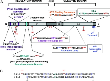
PKCdelta (protein kinase Cdelta) is a serine/threonine kinase that plays a key role in growth regulation and tissue remodelling. Traditional models of PKC activation have focused on lipid cofactors and anchoring proteins that localize the active conformation of PKCdelta to membranes, in close proximity with its target substrates. However, recent studies identify a distinct mode for PKCdelta activation involving tyrosine phosphorylation by Src family kinases. The tyrosine-phosphorylated form of PKCdelta (which accumulates in the soluble fraction of cells exposed to oxidant stress) displays lipid-independent kinase activity and is uniquely positioned to phosphorylate target substrates throughout the cell (not just on lipid membranes). This review summarizes (1) recent progress towards understanding structure-activity relationships for PKCdelta, with a particular focus on the stimuli that induce (and the distinct functional consequences that result from) tyrosine phosphorylation events in PKCdelta's regulatory, hinge and catalytic domains; (2) current concepts regarding the role of tyrosine phosphorylation as a mechanism to regulate PKCdelta localization and actions in mitochondrial and nuclear compartments; and (3) recent literature delineating distinct roles for PKCdelta (relative to other PKC isoforms) in transcriptional regulation, cell cycle progression and programmed cell death (including studies in PKCdelta-/- mice that implicate PKCdelta in immune function and cardiovascular remodelling). Collectively, these studies argue that the conventional model for PKCdelta activation must be broadened to allow for stimulus-specific differences in PKCdelta signalling during growth factor stimulation and oxidant stress.
Figures
References
-
- Dempsey E. C., Newton A. C., Mochly-Rosen D., Fields A. P., Reyland M. E., Insel P. A., Messing R. O. Protein kinase C isozymes and the regulation of diverse cell responses. Am. J. Physiol. Lung Cell. Mol. Physiol. 2000;279:L429–L438. - PubMed
-
- Sabri A., Steinberg S. F. Protein kinase C isoform-selective signals that lead to cardiac hypertrophy and the progression of heart failure. Mol. Cell. Biochem. 2003;251:97–101. - PubMed
-
- Solaro R. J., Burkart E. M. Functional defects in troponin and the systems biology of heart failure. J. Mol. Cell. Cardiol. 2002;34:689–693. - PubMed
-
- Cho W. Membrane targeting by C1 and C2 domains. J. Biol. Chem. 2001;276:32407–32410. - PubMed
Publication types
MeSH terms
Substances
Grants and funding
LinkOut - more resources
Full Text Sources
Other Literature Sources
Molecular Biology Databases
Miscellaneous