. 2004 Oct 25;167(2):245-56.
doi: 10.1083/jcb.200406008. Epub 2004 Oct 18.

The forces that position a mitotic spindle asymmetrically are tethered until after the time of spindle assembly

Affiliations

The forces that position a mitotic spindle asymmetrically are tethered until after the time of spindle assembly

Jean-Claude Labbé et al. J Cell Biol. .

Abstract

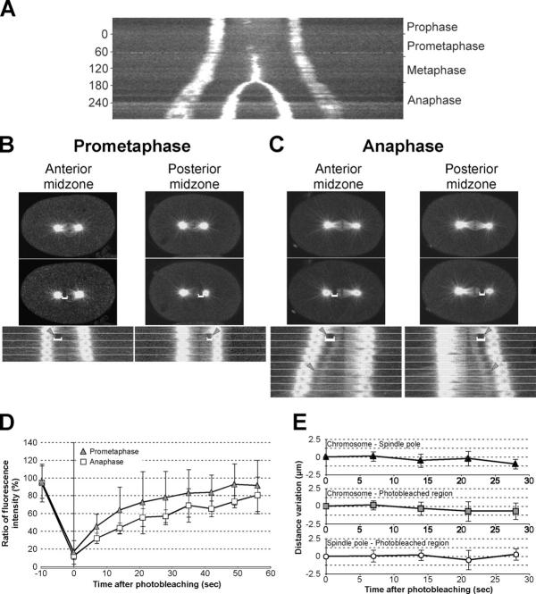
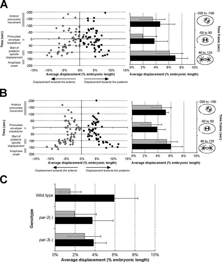
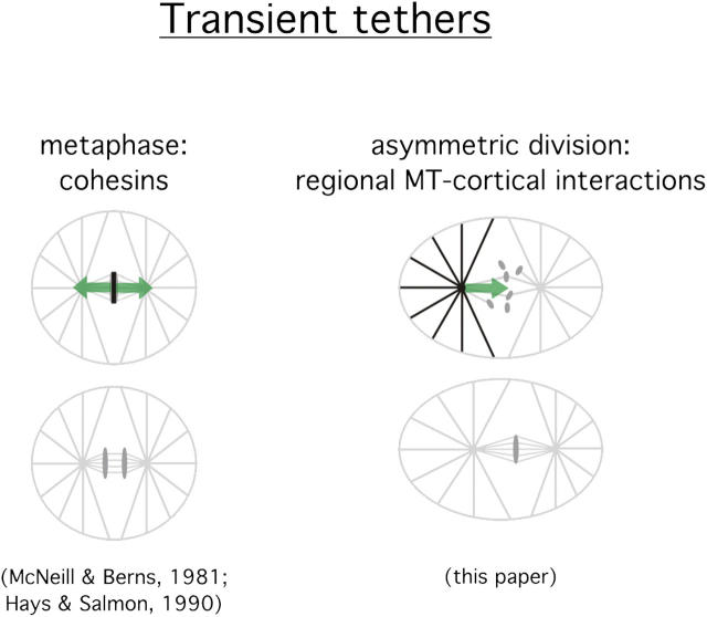
Regulation of the mitotic spindle's position is important for cells to divide asymmetrically. Here, we use Caenorhabditis elegans embryos to provide the first analysis of the temporal regulation of forces that asymmetrically position a mitotic spindle. We find that asymmetric pulling forces, regulated by cortical PAR proteins, begin to act as early as prophase and prometaphase, even before the spindle forms and shifts to a posterior position. The spindle does not shift asymmetrically during these early phases due to a tethering force, mediated by astral microtubules that reach the anterior cell cortex. We show that this tether is normally released after spindle assembly and independently of anaphase entry. Monitoring microtubule dynamics by photobleaching segments of microtubules during anaphase revealed that spindle microtubules do not undergo significant poleward flux in C. elegans. Together with the known absence of anaphase A, these data suggest that the major forces contributing to chromosome separation during anaphase originate outside the spindle. We propose that the forces positioning the mitotic spindle asymmetrically are tethered until after the time of spindle assembly and that these same forces are used later to drive chromosome segregation at anaphase.


Figures

Figure 1.
Figure 1.
Posterior spindle displacement begins at metaphase. (A) Time-lapse images of an early C. elegans embryo expressing both γ-tubulin and histone H2B fused to GFP. (B) Kymograph analysis of spindle behavior from these time-lapse images. In both panels, arrowheads point to centrosomes at early metaphase and arrows point to the centrosomes at late metaphase, before anaphase onset. Displacement of the spindle toward the posterior can be observed during metaphase. Displacement began during early metaphase or at the end of prometaphase in all embryos examined in this way (n = 8; see Fig. S1, available at http://www.jcb.org/cgi/content/full/jcb.200406008/DC1). Bars, 5 μm.
Figure 2.
Figure 2.
Posterior spindle displacement occurs in metaphase-arrested embryos. (A) Time-lapse images of untreated wild-type embryos and wild-type embryos treated with c-LβL during pronuclear migration, before meeting. All cellular events appear to occur normally in both cases until anaphase onset. Embryos treated with c-LβL remain arrested in metaphase and fail to undergo cytokinesis. Arrowheads indicate the position of anterior and posterior centrosomes in these time-lapse images. Time is indicated in minutes and the 0 time point is at pronuclear envelope breakdown. Bar, 5 μm. (B) Quantification of the distance between the two spindle poles in untreated (closed circles) and c-LβL–treated (open triangles) wild-type embryos. At the normal time of anaphase onset, the spindle fails to elongate in treated embryos and remains at constant length for at least 30 min. The 0 time point corresponds to pronuclear envelope breakdown. (C) Quantification of the extent of posterior spindle displacement at anaphase onset in untreated and c-LβL–treated, metaphase-arrested wild-type embryos. The spindle initiates posterior movement at the correct time and moves a comparable distance in both cases, and the spindle midpoint was positioned at 56 ± 2% EL in untreated embryos and 56 ± 3% EL in c-LβL–treated embryos. In all panels, n = 5 for untreated embryos and n = 4 for c-LβL–treated embryos. Error bars represent SD over five and four embryos.
Figure 3.
Figure 3.
Laser-mediated disruption of microtubule organization allows the estimation of pulling forces throughout the cell cycle. (A) Two-dimensional reconstruction of multiple confocal sections of embryos fixed and stained for α-tubulin. The centrosomes indicated by arrowheads in the right panels were irradiated before fixation. Laser irradiation specifically disrupts microtubule organization at the targeted centrosome, leaving the unirradiated centrosome largely intact. Bar, 5 μm. (B) Map (left) and quantification (right) of centrosome displacement after OICD. Displacement was determined for anterior centrosomes after posterior centrosome irradiation (gray diamonds and gray bars) and for posterior centrosomes after anterior centrosome irradiation (black squares and black bars). In the right panel, displacements were averaged for various phases of the cell cycle. The timing of cell cycle events was determined according to previously reported values and matches observations made by DIC optics (Labbé et al., 2003). Error bars represent SD over, from top to bottom, 6, 4, 19, 16, 13, and 8 embryos, respectively, for each case. (C) Posterior centrosome movement quantified following no irradiation, laser irradiation of a region between the anterior centrosome and the anterior cortex, or laser irradiation of the whole anterior centrosome. A schematic of the procedure for each condition is shown on the left, with the region irradiated marked with an asterisk. Error bars represent SD over, from top to bottom, 8, 8, and 16 embryos, respectively, for each case. (D) Conceptual model depicting the various types of forces that act on the centrosomes throughout the first mitosis of the early C. elegans embryo. In this model, tethering forces are represented as lines, whereas pulling forces are depicted as springs. At late prophase/prometaphase, the pulling force present in the posterior of the embryo is counteracted by the tethering force in the anterior, thereby preventing posterior spindle displacement. During metaphase, the tethering force in the anterior changes to a pulling force.
Figure 4.
Figure 4.
A transition in forces occurs in metaphase-arrested embryos. Quantification of anterior centrosome movement after posterior centrosome irradiation in untreated (light gray bars) and c-LβL–treated, metaphase-arrested wild-type embryos (dark gray bars). Displacement of the anterior centrosome toward the anterior increases as the cell cycle progresses from late prophase/prometaphase to metaphase. Error bars represent SD over, from top to bottom, 19, 5, 13, 8, 8, and 9 embryos, respectively, for each case.
Figure 5.
Figure 5.
Chromosome segregation occurs without anaphase A and significant poleward flux. (A) Spindle-centered kymograph of an embryo expressing both γ-tubulin and histone H2B fused to GFP. This kymograph was produced similarly to the one in Fig. 1 B, except that posterior movement of the spindle was eliminated: the spindle in each frame of time-lapse images was rotationally aligned and recentered on the midpoint between the centrosomes to allow the observation of symmetries in the spindle. Frames were acquired at 7-s intervals. (B and C) Time-lapse images of embryos expressing β-tubulin::GFP in which a short region of anterior (left) or posterior (right) spindle microtubules were photobleached during prometaphase (B) or anaphase (C) onset. The bottom panels follow FRAP as well as movement of the photobleached region (indicated by gray arrowheads). Frames were acquired at 7-s intervals. For photobleaches of the spindle during prometaphase, kymographs were aligned to the location of chromosomes in the center of the spindle. For photobleaches of the anterior or posterior half of the spindle at anaphase onset, kymographs were aligned to the center of the posterior or anterior centrosome, respectively. (D) Quantification of FRAP during prometaphase (gray triangles) and anaphase (open squares). To correct for fluorophore bleaching and embryo to embryo variations, fluorescence intensity in the photobleached region is expressed as a ratio of bleached over unbleached midzone microtubules in the same embryo. FRAP occurs faster during prometaphase (t 1/2 = 10.6 s; polynomial equation: y = −3E − 07 x4 + 4E − 05x3 − 0.0021x2 + 0.0559x + 0.1571; R2 = 0.997) compared with anaphase (t 1/2 = 17.7 s; polynomial equation: y = 4E − 08x4 + 2E − 06x3 − 0.0005x2 + 0.0303x + 0.1223; R2 = 0.989). Time points were acquired at 7-s intervals. Error bars represent SD over six embryos. (E) Quantification of the distance variation between chromosomes to photobleached region (black triangles), chromosomes and spindle poles (gray squares), and spindle pole to photobleached region (open circles) during anaphase. The distance remains constant between these three positions throughout anaphase. Time points were acquired at 7-s intervals. Error bars represent SD over six embryos.
Figure 6.
Figure 6.
PAR proteins regulate pulling forces throughout the cell cycle. Map (left) and quantification (right) of centrosome displacement after OICD in (A) par-2(lw32) and (B) par-3(it71) mutant embryos. Displacement was determined for anterior centrosomes after posterior centrosome irradiation (gray diamonds and gray bars) and for posterior centrosomes after anterior centrosome irradiation (black squares and black bars). (C) Quantification of anterior (gray bars) and posterior (black bars) centrosome displacement at the time of late prophase/prometaphase in wild-type, par-2(lw32), and par-3(it71) embryos. Error bars represent SD over, from top to bottom, 19, 16, 21, 25, 21, and 21 embryos, respectively, for each case. The overall displacement in par-2 mutant embryos (3.0 ± 2.1% EL, n = 46) was different from wild-type anterior (1.5 ± 1.1% EL, n = 19, P = 0.007) as well as from wild-type posterior (5.9 ± 2.4% EL, n = 16, P = 1.3E−5) centrosomes, and the same was observed when comparing displacement in par-3 mutant embryos (3.4 ± 1.5% EL, n = 42) with that of wild-type anterior (1.5 ± 1.1% EL, n = 19, P = 1.4E−5) and wild-type posterior (5.9 ± 2.4% EL, n = 16, P = 5.3E−6) centrosomes.
Figure 7.
Figure 7.
Model depicting the transient tethers that act during mitosis in cells that divide symmetrically and early C. elegans embryos, which divide asymmetrically. In this model, the net pulling vectorial forces are depicted as arrows and tethers are depicted in dark. In cells that divide symmetrically, tension is present at the kinetochores during metaphase and forces are kept at equilibrium by cohesins that link sister chromatids (McNeill and Berns, 1981; Hays and Salmon, 1990). Cohesins are degraded at anaphase onset, allowing chromosome segregation to occur. In asymmetrically dividing C. elegans embryos, a posterior pulling force is present during late prophase and prometaphase, and this force is kept at equilibrium by the tethering of astral microtubules at the anterior of the embryo. This tether is released during metaphase, allowing posterior movement of the spindle. MT, microtubule.

Similar articles

Cited by

References

    1. Aist, J.R., and M.W. Berns. 1981. Mechanics of chromosome separation during mitosis in Fusarium (Fungi imperfecti): new evidence from ultrastructural and laser microbeam experiments. J. Cell Biol. 91:446–458. - PMC - PubMed
    1. Albertson, D.G. 1984. Formation of the first cleavage spindle in nematode embryos. Dev. Biol. 101:61–72. - PubMed
    1. Brust-Mascher, I., and J.M. Scholey. 2002. Microtubule flux and sliding in mitotic spindles of Drosophila embryos. Mol. Biol. Cell. 13:3967–3975. - PMC - PubMed
    1. Cleveland, D.W., Y. Mao, and K.F. Sullivan. 2003. Centromeres and kinetochores: from epigenetics to mitotic checkpoint signaling. Cell. 112:407–421. - PubMed
    1. Colombo, K., S.W. Grill, R.J. Kimple, F.S. Willard, D.P. Siderovski, and P. Gönczy. 2003. Translation of polarity cues into asymmetric spindle positioning in Caenorhabditis elegans embryos. Science. 300:1957–1961. - PubMed

Publication types

MeSH terms

Substances