. 2004 Oct 18;200(8):967-77.
doi: 10.1084/jem.20040973.

Plasma cell ontogeny defined by quantitative changes in blimp-1 expression

Affiliations

Plasma cell ontogeny defined by quantitative changes in blimp-1 expression

Axel Kallies et al. J Exp Med. .

Abstract

Plasma cells comprise a population of terminally differentiated B cells that are dependent on the transcriptional regulator B lymphocyte--induced maturation protein 1 (Blimp-1) for their development. We have introduced a gfp reporter into the Blimp-1 locus and shown that heterozygous mice express the green fluorescent protein in all antibody-secreting cells (ASCs) in vivo and in vitro. In vitro, these cells display considerable heterogeneity in surface phenotype, immunoglobulin secretion rate, and Blimp-1 expression levels. Importantly, analysis of in vivo ASCs induced by immunization reveals a developmental pathway in which increasing levels of Blimp-1 expression define developmental stages of plasma cell differentiation that have many phenotypic and molecular correlates. Thus, maturation from transient plasmablast to long-lived ASCs in bone marrow is predicated on quantitative increases in Blimp-1 expression.


Figures
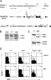
Figure 1.
Figure 1.
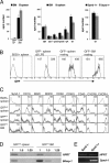
Generation of Blimpgfp reporter mice. (A) The genomic locus of Blimp-1, indicating the exons as boxes and introns as black lines. Coding regions are in gray and nontranslated regions are white. SpeI sites used for Southern hybridization along with the 3′ probe are marked. Arrows indicate direction of translation from initial methionine. The targeted allele derived from the homologous recombination event is indicated. pA, polyadenylation signal sequence; circles, frt sites; triangles, stop codon; splice acc., splice acceptor. The targeted allele encodes a truncated Blimp-1 protein (Blimptrunc) lacking exons 7–8 and GFP from the same mRNA transcript. (B) Southern hybridization of SpeI digested Blimp gfp/+ and C57BL/6 ES cell DNA. (C) Western blot analysis of wild-type and Blimp gfp/+ splenic B cells cultured for 4 d in LPS. The wild-type and Blimptrunc proteins are indicated. β-actin and ICSBP-specific antibodies were used as loading controls. (D) Flow cytometric analysis of spleen and BM. A small population of GFP+ cells was present specifically in the Blimp gfp/+ mice and lost in wild-type or Rag2 −/− Blimp gfp/+ mice.
Figure 2.
Figure 2.
All GFP+ cells are ASC. (A, left) 105 GFP or 200 GFP+ cells from BM or spleen of Blimp gfp/+ mice were assayed for Igκ secretion by ELISPOT. Middle panel, GFP+ cells were sorted from the BM and spleen of Blimp gfp/+ mice and subjected to ELISPOT assay for the indicated isotype. (right) GFP+Synd-1+ and GFP+Synd-1 cells were sorted from BM of Blimp gfp/+ mice assayed for Igκ secretion. Mean number of ELISPOT per 200 cells ±SE is shown. (B) Gated populations show the level of GFP in splenic B220+ B cells and GFP+ ASC from spleen (LPS induced and resting) and BM. GFP+ cells left of the dividing line were considered GFP intermediate (GFPint) and those to the right were considered GFP high (GFPhi). (C) Analysis of the surface phenotype of gated cells from B. Mean fluorescence index is indicated for each histogram. (D) Sorted GFPint and GFPhi cells were sorted as for B and subjected to semi-quantitative RT-PCR analysis. Serial fivefold dilutions of the cDNA were analyzed. (E) BM GFP+Synd-1+ and GFP+Synd-1 cells were sorted and assayed for Synd-1 mRNA. HPRT was a loading control.
Figure 3.
Figure 3.
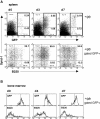
In vivo induction of GFP+ ASC. (A) Blimp gfp/+ mice were injected with 2 μg LPS and analyzed for Synd-1, GFP, and B220 at the indicated time points. (top) Total splenic cells. (bottom) Gated GFP+ cells only. Gated regions are outlined by thick lines. Percentages of GFP+ cells are indicated. (B) Gated GFP+ cells from the same mice were analyzed for relative GFP fluorescence and B220 expression.
Figure 4.
Figure 4.
Development of distinct ASC populations after T cell–dependent immunization. (A) Fluorescence levels of GFP+ cells in spleen, blood, and BM from representative Blimp gfp/+ mice at the indicated times after a single i.p. immunization with NP-KLH in alum. (B) Percentage of GFP+ spleen and BM cells at the indicated time points after immunization. Each circle is an individual mouse. (C) Kinetics of the appearance of GFPint and GFPhi populations in spleen. (D) Frequency of anti-NP IgG1 ASC in GFPint and GFPhi populations in spleen at day 7 (shaded) and day 14 (unshaded) after immunization. Data are from sorted GFP+ cells from three individuals at day 7 and two at day 14.
Figure 5.
Figure 5.
GFPint cells are short-lived plasmablasts. Blimp gfp/+ mice were given a bolus of BrdU and fed BrdU in the drinking water for 4 d. Cells were sorted for the indicated GFP levels from spleen and BM and fixed, and the incorporation of BrdU for each sample was determined by flow cytometry using a BrdU-specific antibody. Data are from two pooled individuals and are representative of three experiments.
Figure 6.
Figure 6.
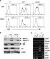
Induction of ASC differentiation in vitro requires Blimp-1. (A) Wild-type, Blimp gfp/+, and Blimp gfp/gfp B cells were cultured in the presence of LPS or the combination CD40L/IL-4/IL-5 for 4 d and examined for GFP expression. Percentages of GFP+ cells are shown. (B) Western blot analysis of splenic B cells cultured in LPS and sorted according to GFP expression. Wild-type cells (WT) are unsorted; (+) GFP+; (−) GFP. The Blimp-1 proteins and the membrane-bound (μM) and secreted IgM (μS) are indicated. Detection of ICSBP was a loading control. White lines indicate intervening lanes have been spliced out. (C) Cells sorted as in B were subjected to RT-PCR analysis for the indicated genes. Blimp-1 primers span exons 7–8 (not expressed from the targeted allele). Blimp gfp/gfp cells do not initiate the ASC transcriptional cascade and remain indistinguishable from the GFP cells. HPRT was used to normalize the relative cDNA input.
Figure 7.
Figure 7.
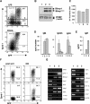
Blimp gfp identifies a heterogeneous population of ASC in vitro. (A) Blimp gfp/+ B cells were stimulated with LPS or CD40L/IL-4/IL-5 for 4 d and examined for Synd-1 and GFP. Four populations GFPSynd-1 (fraction 1), GFP+Synd-1 (fraction 2), GFP+Synd-1+ (fraction 3), and GFPSynd-1+ (fraction 4) cells were sorted and assayed as follows. (B) Western blotting for Blimp-1 protein. ICSBP and β-actin were used as loading controls. (C) Cell proliferation rates. Sorted cells were pulsed with 3[H]thymidine for 2 h. (D) Ig secretion by ELISPOT. Mean numbers of ELISPOT per 100 cells are shown ±SE. (E) IgM secretion. Sorted cells were recultured for 4 h, and supernatants were analyzed by ELISA. (F) Fractions 2 (GFP+/Synd-1+) and 3 (GFP+/Synd-1) were sorted (left) and recultured for 22 h in LPS before reanalysis (right). (G) RT-PCR analysis of the indicated genes was performed on the sorted groups. HPRT was used to normalize the relative cDNA input.
Figure 8.
Figure 8.
Schematic of plasma cell terminal differentiation based on increasing Blimp-1 expression. Relative function or expression status of several parameters is indicated on an arbitrary scale. Dotted lines indicate hypothetical pathways. Analysis of Pax5, IRF4, and CIITA expression is based in vitro evidence only. *, Synd-1 expression on plasma cells is heterogeneous and context specific (see Discussion).

References

    1. Manz, R.A., S. Arce, G. Cassese, A.E. Hauser, F. Hiepe, and A. Radbruch. 2002. Humoral immunity and long-lived plasma cells. Curr. Opin. Immunol. 14:517–521. - PubMed
    1. Manz, R.A., M. Lohning, G. Cassese, A. Thiel, and A. Radbruch. 1998. Survival of long-lived plasma cells is independent of antigen. Int. Immunol. 10:1703–1711. - PubMed
    1. Angelin-Duclos, C., G. Cattoretti, K.I. Lin, and K. Calame. 2000. Commitment of B lymphocytes to a plasma cell fate is associated with Blimp-1 expression in vivo. J. Immunol. 165:5462–5471. - PubMed
    1. Turner, C.A., Jr., D.H. Mack, and M.M. Davis. 1994. Blimp-1, a novel zinc finger-containing protein that can drive the maturation of B lymphocytes into immunoglobulin-secreting cells. Cell. 77:297–306. - PubMed
    1. Schliephake, D.E., and A. Schimpl. 1996. Blimp-1 overcomes the block in IgM secretion in lipopolysaccharide/anti-mu F(ab′)2-co-stimulated B lymphocytes. Eur. J. Immunol. 26:268–271. - PubMed

Publication types

Substances