Comparative Study
. 2004 Sep;5(3):270-84.
doi: 10.1007/s10162-004-4051-4. Epub 2004 Jun 24.

Differences between the negatively activating potassium conductances of Mammalian cochlear and vestibular hair cells

Affiliations
Comparative Study

Differences between the negatively activating potassium conductances of Mammalian cochlear and vestibular hair cells

Weng Hoe Wong et al. J Assoc Res Otolaryngol. 2004 Sep.

Abstract

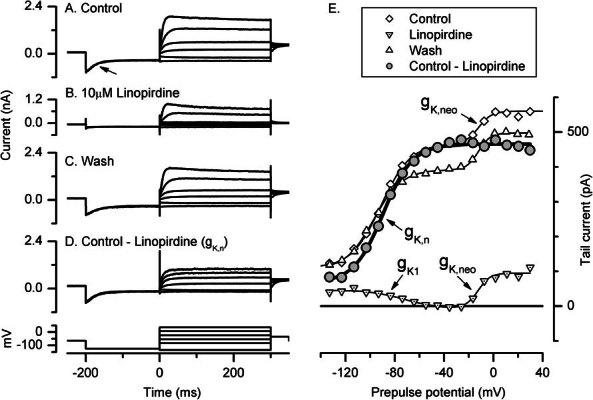
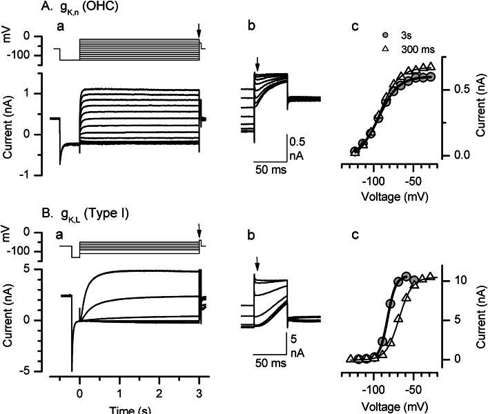
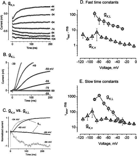
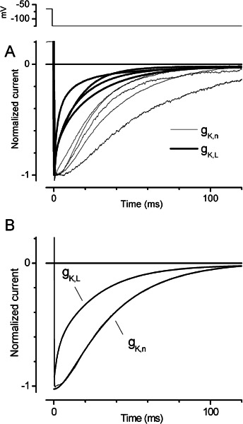
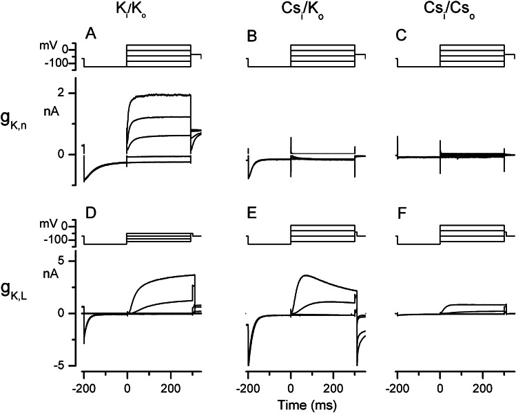
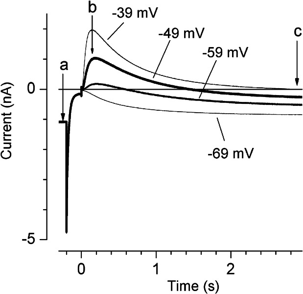
Cochlear and type I vestibular hair cells of mammals express negatively activating potassium (K(+)) conductances, called g(K,n) and g(K,L) respectively, which are important in setting the hair cells' resting potentials and input conductances. It has been suggested that the channels underlying both conductances include KCNQ4 subunits from the KCNQ family of K(+) channels. In whole-cell recordings from rat hair cells, we found substantial differences between g(K,n) and g(K,L) in voltage dependence, kinetics, ionic permeability, and stability during whole-cell recording. Relative to g(K,L), g(K,n) had a significantly broader and more negative voltage range of activation and activated with less delay and faster principal time constants over the negative part of the activation range. Deactivation of g(K,n) had an unusual sigmoidal time course, while g(K,L) deactivated with a double-exponential decay. g(K,L), but not g(K,n), had appreciable permeability to Cs(+). Unlike g(K,L), g(K,n)'s properties did not change ("wash out") during the replacement of cytoplasmic solution with pipette solution during ruptured-patch recordings. These differences in the functional expression of g(K,n) and g(K,L) channels suggest that there are substantial differences in their molecular structure as well.


Figures

Figure 1
Figure 1
gK,n and other voltage-dependent currents in a rat outer hair cell, revealed by their voltage dependence and sensitivity to the KCNQ blocker, linopirdine. Whole-cell currents were recorded from an outer hair cell isolated from the apical turn of a P17 rat cochlea. A–C Whole-cell currents evoked by the voltage protocol shown below, in control solution (high-Ca2+ L15) (A), control + 10 μM linopirdine (B) and wash (high-Ca2+ L15) (C). Voltage protocol shown at the bottom. From the holding potential of −64 mV, voltage was stepped to −124 mV, which in control solutions deactivated gK,n (A,C) (arrow in A), and then to a series of steps between −134 and + 36 mV; gK,n was reactivated by steps positive to −124 mV. At the offset of the iterated voltage steps, the voltage was stepped to −34 mV and peak tail currents were measured. These currents, which reflect the activation state of the channels at the end of the preceding voltage steps, are plotted as functions of the prepulse voltage in E. Linopirdine (B) blocked the current component that deactivated during the hyperpolarizing step (arrow in A), and much of the current during the iterated voltage steps. D. The linopirdine-blocked current (through gK,n), obtained by subtracting records in B from records in A. E. Activation curves generated from tail currents at the offset of the iterated voltage steps; the tail current voltage was −34 mV. In control and wash, the curves are fit by the sum of two Boltzmanns [Eq.(2)], with V1/2 and S values of −91.3 mV and 11.9 mV, respectively, and −12.0 mV and 5.3 mV (control data). Linopirdine fully blocked gK,n, partly blocked (see text) an inwardly rectifying conductance (gK1 ; V1/2 = −73.5 mV, S = 11.3 mV), and did not affect the more positively activating outward rectifier (gK,neo, V1/2 = −13.25 mV, S = 3.7 mV). The linopirdine-sensitive activation curve (gK,n, from D) is well fit by a single Boltzmann with V1/2 = −89.4 mV and S = 12.2 mV.
Figure 2
Figure 2
Differences in the voltage dependences of gK,n and gK,L. Whole-cell currents were recorded from a P16 outer hair cell (Aa) and a P17 type I cell (Ba) with 3-s voltage step protocols (top panels). Aa. Steps to −124 mV from a holding potential of −64 mV deactivated gK,L. Ba. Steps to −129 mV from a holding potential of −69 mV deactivated gK,L. Depolarizing steps reactivated the conductances. Ab, Bb. At the offset of the iterated voltage steps, the voltage was stepped to −34 mV (Aa) and −39 mV (Ba) and tail currents were measured (at the arrows) for plotting against prepulse voltage in c. Ac, Bc. Activation curves for the tail current data shown in Ab,Bb (filled circles) are fit with a Boltzmann function [Eq.(1) thick lines, yielding V1/2 and S values of −93. 6 mV and 12.4 mV (AC, gK,n) and −82.4 mV and 5.0 mV (Bc, gK,L), near the mean values for our samples of outer hair cells and type I hair cells, respectively. In this outer hair cell, unlike the cell in Figure 1, the activation curve is well fit by a single Boltzmann, with no indication of a second outward rectifier at depolarized potentials. This is not related to the long-duration iterated voltage steps, as a similar activation curve was obtained with 300-ms voltage steps (Ac, (triangles and thin line: V1/2 and S values of −92.8 mV and 12.3 mV). In the type I cell, in contrast, the activation curve taken after 300-ms voltage steps (triangles) was positively shifted (thin line): V1/2 = −68.1 mV and S = 8.19 mV.
Figure 3
Figure 3
Differences in the activation kinetics of gK,n and gK,L. A,B. Fitting of activation kinetics with Eq.(3) for (A) gK,n from a P16 outer hair cell and (B) gK,L from a P19 type I hair cell. Voltage was first stepped to −124 mV for 500 ms (A) and −129 mV for 200 ms (B) to fully deactivate the conductances, then stepped to the values indicated by each trace. gK,n: V1/2= −87.5, S = 14.1 mV, no second outward rectifier, average of 3 traces at each voltage; gK,L: V1/2 = −83.6, S = 5.0 mV, not averaged. C. Currents from A and B at two similar command voltages, normalized to the current at 3 s for comparison of kinetics. gK,L activated much more slowly at voltages near −90 mV and with a longer delay at voltages near −50 mV. Note that the reversal potentials for these K+ conductances were approximately −80 mV, so that current was inward at −90 mV and outward at −50 mV. D,E. Fast (D) and slow (E) time constants as functions of voltage. Triangles values for gK,n averaged from 5 OHCs. Circles, values for gK,L averaged from 4–5 type I hair cells, except for the data points at −29 mV, which are averages of fits from two cells (τslow = 13.4, 27.2 ms; τfast = 13.4, 11.9ms).
Figure 4
Figure 4
Differences in the deactivation kinetics of gK,n and gK,L. gK,L deactivation followed a single- or double-exponential decay function, while gK,n deactivation had a sigmoidal onset. A. Steps from −64 to −124 mV (gK,n) or -69 to −129 mV (gK,L) caused deactivation of currents through both gK,n, and gK,L ; thin traces are for gK,n from 5 outer hair cells, thick traces are for gK,L from 5 type I cells. Currents are normalized to the peak current at the beginning of the step. B. Fits (thick lines) to the deactivation of two of the data traces in A (thin lines; obscured by fits except at the step onset). The type I data were fit by a double-exponential function [eq.(4)]: A1 = −264 pA, τ1 = 2.5 ms, A2 = −684 pA, τ2 = 26.8 ms. The OHC data were fit by Eq.(3): τ1 = 7.8 ms, τ2 = 33.1 ms.
Figure 5
Figure 5
gK,L , but not gK,n, is Cs+ permeable. Left column: Currents in control conditions, with high internal K+ and 5.8-mM external K+. Middle column (different cells): With an internal Cs+ solution, gK,n (B) carried only inward current but gK,L (E) still passed both inward and outward current. Right column (same cells as middle column): When, in addition, external K+ was replaced with Cs+, all current through gK,n was blocked (C; residual slope conductance = 600 pS) but outward current still flowed in the type I cell (F). Current and voltage axes on the left apply to all panels in a row. In standard solutions, relatively small voltage steps applied to the type I cell (D) elicited very large currents through gK,L. Currents are from a P16 OHC (A), a P17 OHC (B,C), a P24 type I cell (D), and a P23 type I cell (E,F).
Figure 6
Figure 6
Changes in the reversal potential of gK,L during long voltage steps may result from significant K+ flux through gK,L. Standard vestibular external medium and Cs+ pipette solution for perforated patch recordings. Same cell as in Figure 5B,C. After a 200-ms step to −129 mV, the peak currents (b, arrow) reversed at approximately -65 mV. With time during 3-s steps positive to −69 mV, however, the reversal potential shifted positively, reaching approximately −40 mV at the end of the steps (c). The change in reversal potential is shown clearly by the reversal of current polarity during the steps at −59 and −49 mV (thick traces).

Similar articles

Cited by

References

    1. Angelaki DE. Three-dimensional organization of otolith-ocular reflexes in rhesus monkeys. III. Responses to translation. J. Neurophysiol. 1998;80:680–695. - PubMed
    1. Angelo K, Jespersen T, Grunnet M, Nielsen MS, Klaerke DA, Olesen SP. KCNE5 induces time- and voltage-dependent modulation of the KCNQ1 current. Biophys. J. 2002;83:1997–2006. - PMC - PubMed
    1. Art JJ, Fettiplace R. Variation of membrane properties in hair cells isolated from the turtle cochlea. J Physiol. 1987;385:207–242. - PMC - PubMed
    1. Ashmore JF. Forward and reverse transduction in the mammalian cochlea. Neurosci. Res. Suppl. 1990;12:S39–S50. doi: 10.1016/0921-8696(90)90007-P. - DOI - PubMed
    1. Barry PH. JPCalc, a software package for calculating liquid junction potential corrections in patch-clamp, intracellular, epithelial and bilayer measurements and for correcting junction potential measurements. J. Neurosc. Meth. 1994;51:107–116. doi: 10.1016/0165-0270(94)90031-0. - DOI - PubMed

Publication types