. 2004 Oct 22;16(2):199-209.
doi: 10.1016/j.molcel.2004.09.021.

Global position and recruitment of HATs and HDACs in the yeast genome

Affiliations

Global position and recruitment of HATs and HDACs in the yeast genome

François Robert et al. Mol Cell. .

Abstract

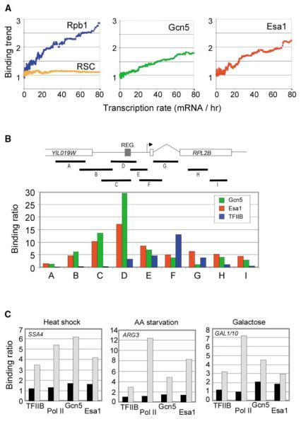
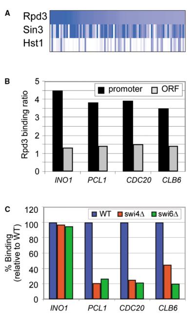
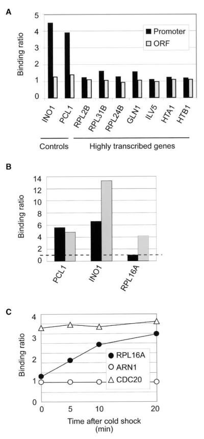
Chromatin regulators play fundamental roles in the regulation of gene expression and chromosome maintenance, but the regions of the genome where most of these regulators function has not been established. We explored the genome-wide occupancy of four different chromatin regulators encoded in Saccharomyces cerevisiae. The results reveal that the histone acetyltransferases Gcn5 and Esa1 are both generally recruited to the promoters of active protein-coding genes. In contrast, the histone deacetylases Hst1 and Rpd3 are recruited to specific sets of genes associated with distinct cellular functions. Our results provide new insights into the association of histone acetyltransferases and histone deacetylases with the yeast genome, and together with previous studies, suggest how these chromatin regulators are recruited to specific regions of the genome.


Figures

Figure 1
Figure 1
Recruitment of Histone Acetylases Gcn5 and Esa1 to Promoters of Actively Transcribed Genes (A) Correlation between chromatin regulator occupancy and transcription rates. The binding trend was calculated by computing the moving median of the binding ratio over a sliding window of 100 genes across all genes ordered by transcription rate as described previously for other regulators such as Set1 (Ng et al., 2002, 2003). The transcription rates for yeast genes and the binding data for RSC were determined previously (Holstege et al., 1998; Ng et al., 2002). Rpb1 occupancy correlates with transcription rate across the genome, whereas RSC occupancy does not, and these proteins served as controls. (B) Fine mapping of Gcn5 (green), Esa1 (red), and TFIIB (blue) occupancy within the RPL2B locus. The binding ratios from segment-specific chromatin IP experiments are shown for Gcn5, Esa1, and TFIIB for each of the DNA segments A–I. The binding ratios (representing the enrichment generated by the ChIP) were all normalized to the promoter of ARN1, which was set to 1. (C) The binding ratios for the general transcription factor TFIIB, RNA pol II (8WG16), Gcn5, and Esa1 are shown in uninduced (black) and induced (gray) states for heat shock (25°C versus 15 min at 37°C) at the SSA4 promoter, amino acid starvation (YNB versus 10 min in minimal synthetic media + sulfometuron methyl) at the ARG3 promoter, and galactose (raffinose versus galactose) at the GAL1/10 promoters. Binding ratios were calculated as in (B). The exact growth and inducing conditions can be found in the Supplemental Data. These experiments were repeated multiple times and the variation was never more than 15%.
Figure 2
Figure 2
Recruitment of Hst1 to Promoters of Sporulation and Kynureine Genes by the Sum1 Repressor (A) The binding ratio for Hst1 (blue), Sum1 (red), and Hst1 in sum1Δ cells (green) across the SPS4 and BNA5 loci, as determined by ChIP, is shown. A diagram of the regions amplified by sequence-specific PCR is shown on top. (B) The log binding ratio (as determined by genome-wide location analysis) of Hst1, Sum1, and Rpd3 (used as a control) is displayed for the promoters of genes bound by Hst1 (p < 0.005). The log binding ratio of Hst1 in sum1 deletion cells (sum1Δ) is also shown. (C) The level of histone H3 (K9/K14) acetylation and histone H4 (K5, K8, K12, K16) acetylation, as determined by ChIP, is shown in wild-type cells (black) and in cells deleted for HST1 (gray) at the SPS4 and BNA5 promoters. The binding ratios in (A) and (C) were all calculated as in Figure 1B. The binding ratios in hst1Δ in (C) were normalized to wt. These experiments were repeated multiple times and the variation was never more than 15%.
Figure 3
Figure 3
Occupancy and Recruitment of Rpd3 to Promoters of Cell Cycle Genes (A) Sin3 is associated with virtually all the genes bound by Rpd3. The log binding ratio (as determined by genome-wide location analysis) of Rpd3, Sin3, and Hst1 (used as a control) is displayed for the promoters of genes bound by Rpd3 (p < 0.005). (B) Rpd3 is associated with the promoters but not the open reading frames (ORFs) of target genes. The Rpd3 binding ratio, as determined by gene-specific ChIP, is shown for the promoter (black) and ORF (gray) of selected Rpd3 target genes. (C) The association of Rpd3 with promoters of PCL1, CDC20, and CLB6 is dependent on Swi4 and Swi6. The binding ratio of Rpd3 is shown for various Rpd3 target promoters in wild-type cells (blue) and in cells deleted for SWI4 (red) or SWI6 (green). The binding ratios in (B) and (C) were all calculated as in Figure 1B. These experiments were repeated multiple times and the variation was never more than 15%.
Figure 4
Figure 4
Rpd3 Is Recruited to Ribosomal Protein Genes upon Cold Shock (A) Rpd3 is not generally associated with highly transcribed protein-coding genes in rich media. Gene-specific ChIPs were performed with an Rpd3-tagged strain at the promoter (black) and ORF (gray) of the indicated highly transcribed genes. INO1 and PCL1 were included as positive controls. (B) Different protocols give different enrichment at ribosomal protein genes. Rpd3 ChIP experiments were performed using our standard protocol (black) and the Kurdistani et al. (2002) protocol (gray). The binding ratio of Rpd3 is shown at the INO1, PCL1, and RPL16A promoters. These experiments were repeated multiple times and the variation was never more than 15%. (C) Cold shock treatment induces the binding of Rpd3 to ribosomal protein genes. Myc-Rpd3 cells were grown until mid-log phase and the culture was transferred to an ice-cold water bath. 50 ml samples were collected and subject to ChIP prior to, as well as 5, 10, and 20 min after transferring to cold water bath. The binding of Rpd3 is shown at the CDC20, ARN1 (a negative control), and RPL16A promoters. The binding ratios were all calculated as in Figure 1B. These experiments were repeated multiple times and the variation was never more than 15%.

References

    1. Allard S, Utley RT, Savard J, Clarke A, Grant P, Brandl CJ, Pillus L, Workman JL, Cote J. NuA4, an essential transcription adaptor/histone H4 acetyltransferase complex containing Esa1p and the ATM-related cofactor Tra1p. EMBO J. 1999;18:5108–5119. - PMC - PubMed
    1. Allfrey VG, Faulkner R, Mirsky AE. Acetylation and methylation of histones and their possible role in the regulation of RNA synthesis. Proc. Natl. Acad. Sci. USA. 1964;51:786–794. - PMC - PubMed
    1. Arevalo-Rodriguez M, Cardenas ME, Wu X, Hanes SD, Heitman J. Cyclophilin A and Ess1 interact with and regulate silencing by the Sin3-Rpd3 histone deacetylase. EMBO J. 2000;19:3739–3749. - PMC - PubMed
    1. Baek SH, Ohgi KA, Rose DW, Koo EH, Glass CK, Rosenfeld MG. Exchange of N-CoR corepressor and Tip60 coactivator complexes links gene expression by NF-kappaB and beta-amyloid precursor protein. Cell. 2002;110:55–67. - PubMed
    1. Bannister AJ, Zegerman P, Partridge JF, Miska EA, Thomas JO, Allshire RC, Kouzarides T. Selective recognition of methylated lysine 9 on histone H3 by the HP1 chromo domain. Nature. 2001;410:120–124. - PubMed

Publication types

MeSH terms