Comparative Study
. 2004 Dec 24;279(52):54808-16.
doi: 10.1074/jbc.M406654200. Epub 2004 Oct 19.

The GAT domains of clathrin-associated GGA proteins have two ubiquitin binding motifs

Affiliations
Comparative Study

The GAT domains of clathrin-associated GGA proteins have two ubiquitin binding motifs

Patricia S Bilodeau et al. J Biol Chem. .

Abstract

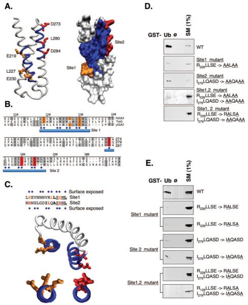
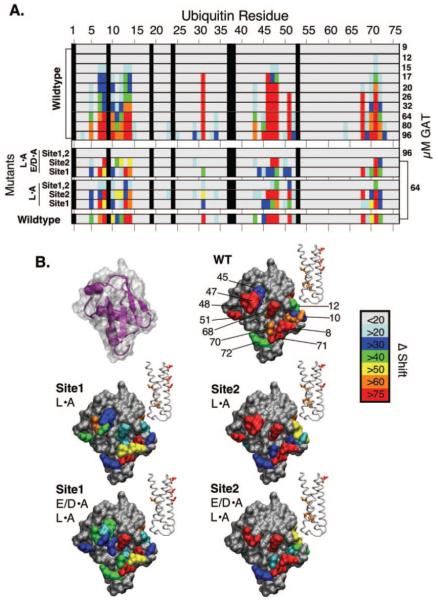
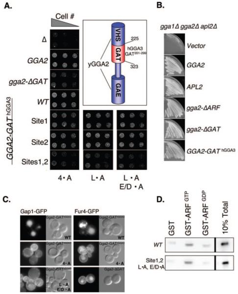
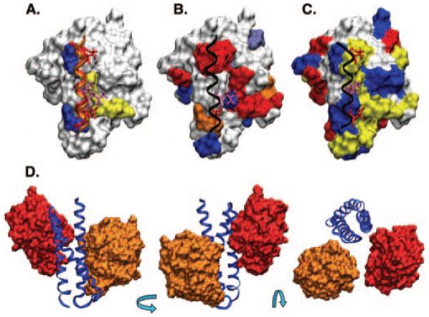
Ubiquitin (Ub) attachment to membrane proteins can serve as a sorting signal for lysosomal delivery. Recognition of Ub as a sorting signal can occur at the trans-Golgi network and is mediated in part by the clathrin-associated Golgi-localizing, gamma-adaptin ear domain homology, ARF-binding proteins (GGA). GGA proteins bind Ub via a three-helix bundle subdomain in their GAT (GGA and target of Myb1 protein) domain, which is also present in the Ub binding domain of target of Myb1 protein. Ubiquitin binding by yeast Ggas is required to direct sorting of ubiquitinated proteins such as general amino acid permease (Gap1) from the trans-Golgi network to endosomes. Using affinity chromatography and nuclear magnetic resonance spectroscopy, we have found that the human GGA3 GAT domain contains two Ub binding motifs that bind to the same surface of ubiquitin. These motifs are found within different helices within the three-helix GAT subdomain. When functionally analyzed in yeast, each motif was sufficient to mediate trans-Golgi network to endosomal sorting of Gap1, and mutation of both motifs resulted in defective Gap1 sorting without defects in other GGA-dependent processes.


Figures

Fig. 1
Fig. 1. Human GGA3 has two conserved ubiquitin binding motifs
A, a predicted model of the C-terminal three-helix bundle of the human GGA3 GAT domain. Shown in blue are two regions conserved among other ubiquitin-binding proteins with GAT domains. Also shown are residues proposed to comprise Ub binding interfaces. Residues in Site1 are present on the N-terminal α-helix and are colored orange. Residues in Site2 are red. B, an alignment of GAT domains from human GGA3, Tom1, and yeast GGA2. The numbering corresponds to the residue position of the long form of hGGA3. The two conserved regions in blue in panel A are underscored with a blue bar. Residues predicted to be on the same external face of an α-helix are designated with a dot. C, sequences of two putative Ub binding motifs are aligned at the top. Predicted surface residues are designated with a dot. Middle, the region of the GAT domain with both Ub binding motifs. Bottom, views of each α-helical region showing the alignment of Glu-219, Leu-227, and Glu-230 as well as Asp-273, Leu-280, and Asp-284 on the external face of the helix. D, bacterial lysates containing wild-type or mutant GAT domains were incubated with 100 μl of GSH-agarose bound with GST alone (ø) or GST·Ub (Ub). Beads were washed and immunoblotted together with the indicated fraction of the starting lysate with anti-V5 antibodies directed to an N-terminal hexohistidine/V5 epitope tag. GAT domains with mutations in Site1 and/or Site2 are underlined. E, wild-type and mutant GAT domains were purified over nickel-agarose and incubated (10 μg) with 100 μl of GSH-agarose bound with GST alone (ø) or GST·Ub (Ub). Beads were washed and immunoblotted together with the indicated fraction of the starting lysate with anti-V5 antibodies. GAT domains with mutations in Site1 and/or Site2 are underlined.
Fig. 2
Fig. 2. Both GAT ubiquitin binding motifs bind the same surface
A, the HSQC spectra of 15N-labeled Ub (110 μm) were measured in the presence of increasing quantities of wild-type GAT domain (shown on right) as well as in the presence of mutant GAT domains. The chemical shift differences induced by GAT binding were quantified using (0.2 ∂N2 + ∂H2)1/2, and values were normalized using the maximal shift change observed with wild-type GAT domain at 64 μm. The magnitude of chemical shift change was then plotted colorimetrically for each residue using the color scale indicated. Each mutant GAT domain was measured at 64 μm except for the one indicated mutant measured at 96 μm. GAT domain mutants with a single alanine substitution of Leu-227 (Site1) and/or Leu-280 (Site2) are designated as L·A. Mutants with double mutations in Site 1 (L227A,E230A) and/or Site2 (L280A,D284A) are designated L·A, E/D ·A. B, using the color scale in panel A, residues undergoing chemical shift changes were plotted onto the surface of Ub. Also pictured (upper right) is the secondary structure of Ub overlaid with the surface. The structure of corresponding GAT domains that induced chemical shift changes on each Ub surface are modeled in the inset. Mutant GAT domains containing single or double alanine substitutions in either Site1 or Site2 follow the same nomenclature and are designated L·A or L·A, E/D·A, respectively.
Fig. 3
Fig. 3. Elimination of both ubiquitin binding motifs results in ADCB-sensitive growth
A, cells containing the designated yeast GGA2 alleles as their sole GGA gene were serially diluted and plated onto synthetic media containing ammonia as the nitrogen source and 75 μm ADCB. Chimeras using an HA epitope-tagged GGA2 were made in which the C-terminal three-helix bundle region of GGA2 (residues 226–322) was replaced with the corresponding region of either wild-type hGGA3 (residues 201–299) or mutant hGGA3 to make a series of GGA2-hGGAGAT alleles. A schematic of these alleles is shown in the inset. The GGA2 and gga2GAT alleles were integrated into the genome; other alleles were carried on low copy plasmids. GAT domain mutants with a single alanine substitution of Leu-227 (Site1) and/or Leu-280 (Site2) are designated as L·A. Mutants with double mutations in Site 1 (L227A,E230A) and/or Site2 (L280A,D284A) are designated as L·A, E/D ·A. GAT domain mutants carrying four alanine substitutions in Site1 and/or Site2 are designated as 4·A. B, triple mutant cells (gga1Δ,gga2Δ,apl2Δ) carrying a URA3-based plasmid with wild-type GGA2 were transformed with a low copy LEU2-based plasmid carrying wild-type GGA2, APL2, or the indicated gga2 or GGA2-hGGA3GAT alleles. Cells were then plated onto 5′-fluoroorotic acid to select for cells that could tolerate loss of the URA3-based GGA2. C, cells (ggaΔ end3Δ) carrying the gga2GAT allele or low copy plasmids expressing GGA2-hGGA3GAT without or with the indicated mutations in both Ub binding Sites were transformed with plasmids expressing Gap1·GFP or Fur4·GFP under the copper-inducible control of the CUP1 promoter. Cells were grown overnight in the absence of copper, pelleted, and resuspended in the presence of 200 μm copper. For Gap1·GFP sorting, cells were grown in YPD for 4 h prior to viewing. For Fur4·GFP sorting, cells were grown in 50 μg/ml excess uracil for 7 h prior to viewing. D, wild-type hGGA3 GAT domain and the Sites1,2 mutant GAT domain (L227A,E230A,L280A,D284A) were incubated with GST, GST·ARFGTP, or GST·ARFGDP. Beads were washed and immunoblotted with anti-V5 antibodies recognizing an N-terminal V5 epitope on each GAT domain.
Fig. 4
Fig. 4. Model for ubquitin interaction with the GAT domain
A, the relevant helical portion encompassing Site1 and Site2 of the GAT domain docked with Ub. The hydrophobic patch (Leu-8, Ile-44, and Val-70) of Ub shown in yellow and basic residues of Ub (Lys-48 and Arg-72) in blue are coordinated with the central leucine and flanking acidic residues of the two GAT binding Sites, respectively. The side chains of Site1 (219, 227 230) and Site2 (273, 280, 284) are shown. B, the helical portion of Site1 with only Ub is shown where the surface of Ub is color-coded according to the chemical shift perturbations quantified in Fig. 2 using the wild-type hGGA3 domain and 15N-Ub. C, the helical portion of Site2 with only Ub where the surface of Ub is color-coded for acidic residues (red), basic residues (blue), and non-polar residues (yellow). D, views of a single hGGA3 GAT domain (blue) with both Ub binding Sites bound to Ub. Ub bound to Site1 is in orange; Site2 is in red.

Similar articles

Cited by

References

    1. Hicke L, Dunn R. Annu. Rev. Cell Dev. Biol. 2003;19:141–172. - PubMed
    1. Shih SC, Sloper-Mould KE, Hicke L. EMBO J. 2000;19:187–198. - PMC - PubMed
    1. Booth JW, Kim MK, Jankowski A, Schreiber AD, Grinstein S. EMBO J. 2002;21:251–258. - PMC - PubMed
    1. Shih SC, Katzmann DJ, Schnell JD, Sutanto M, Emr SD, Hicke L. Nat. Cell Biol. 2002;4:389–393. - PubMed
    1. Polo S, Sigismund S, Faretta M, Guidi M, Capua MR, Bossi G, Chen H, De Camilli P, Di Fiore PP. Nature. 2002;416:451–455. - PubMed

Publication types

MeSH terms

LinkOut - more resources