. 2004 Oct 19;32(18):5596-608.
doi: 10.1093/nar/gkh863. Print 2004.

The C-terminal alphaO helix of human Ogg1 is essential for 8-oxoguanine DNA glycosylase activity: the mitochondrial beta-Ogg1 lacks this domain and does not have glycosylase activity

Affiliations

The C-terminal alphaO helix of human Ogg1 is essential for 8-oxoguanine DNA glycosylase activity: the mitochondrial beta-Ogg1 lacks this domain and does not have glycosylase activity

K Hashiguchi et al. Nucleic Acids Res. .

Abstract

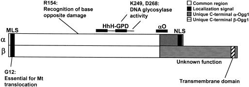
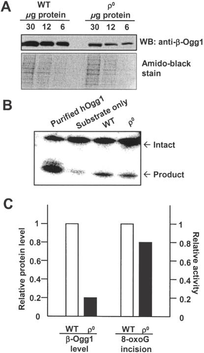
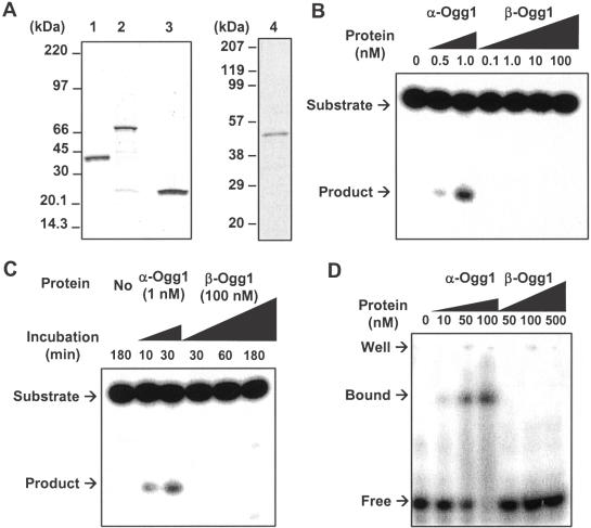
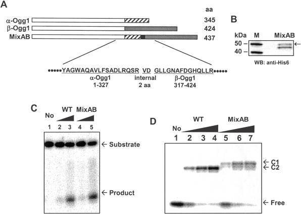
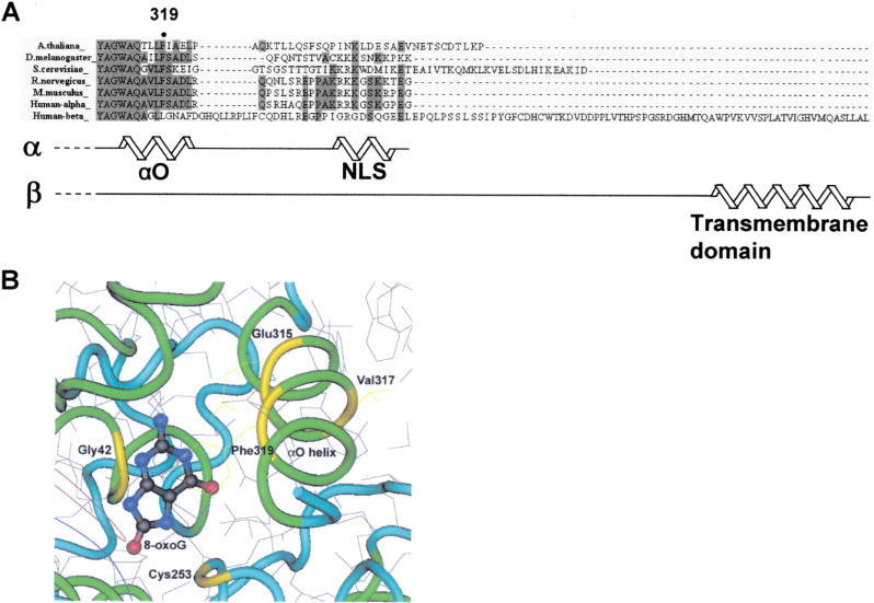
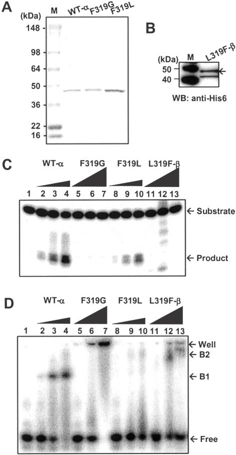
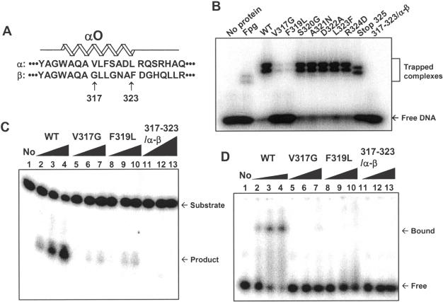
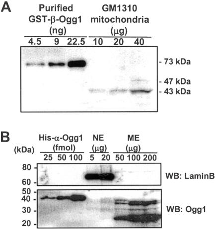
The human Ogg1 glycosylase is responsible for repairing 8-oxo-7,8-dihydroguanine (8-oxoG) in both nuclear and mitochondrial DNA. Two distinct Ogg1 isoforms are present; alpha-Ogg1, which mainly localizes to the nucleus and beta-Ogg1, which localizes only to mitochondria. We recently showed that mitochondria from rho(0) cells, which lack mitochondrial DNA, have similar 8-oxoG DNA glycosylase activity to that of wild-type cells. Here, we show that beta-Ogg1 protein levels are approximately 80% reduced in rho(0) cells, suggesting beta-Ogg1 is not responsible for 8-oxoG incision in mitochondria. Thus, we characterized the biochemical properties of recombinant beta-Ogg1. Surprisingly, recombinant beta-Ogg1 did not show any significant 8-oxoG DNA glycosylase activity in vitro. Since beta-Ogg1 lacks the C-terminal alphaO helix present in alpha-Ogg1, we generated mutant proteins with various amino acid substitutions in this domain. Of the seven amino acid positions substituted (317-323), we identified Val-317 as a novel critical residue for 8-oxoG binding and incision. Our results suggest that the alphaO helix is absolutely necessary for 8-oxoG DNA glycosylase activity, and thus its absence may explain why beta-Ogg1 does not catalyze 8-oxoG incision in vitro. Western blot analysis revealed the presence of significant amounts of alpha-Ogg1 in human mitochondria. Together with previous localization studies in vivo, this suggests that alpha-Ogg1 protein may provide the 8-oxoG DNA glycosylase activity for the repair of these lesions in human mitochondrial DNA. beta-Ogg1 may play a novel role in human mitochondria.


Figures

Figure 1
Figure 1
Structure of the two major human Ogg1 isoforms. Schematics of α- (345 amino acids) and β-Ogg1 (424 amino acids) proteins are shown. The first 316 amino acids are common for both isoforms, while the C-termini vary considerably. The mitochondrial localization signal (MLS, position 9–26), nuclear localization signal (NLS, 335–342) and HhH-GPD motif are indicated. The functions of amino acids Gly-12, Arg-154, Lys-249 and Asp-268 are indicated, based on previous functional studies. The function of the β-Ogg1 C-terminus is unknown. A transmembrane domain (position 400–422) in a highly hydrophobic region was predicted by the SOSUI system (32).
Figure 2
Figure 2
β-Ogg1 protein levels in human mitochondria lacking mtDNA. (A) β-Ogg1 protein levels in mitochondria from WT and ρ0 cells were analyzed by western blot using β-Ogg1 antibody. An amido black-stained PVDF membrane is also presented to show equal loading of protein. (B) Incision of an 8-oxoG-containing substrate by WT and ρ0 mitochondrial extracts was measured as described in Materials and Methods. (C) β-Ogg1 protein levels and incision activity are presented relative to the values observed with WT cells. 8-oxoG DNA glycosylase activity was measured in our previous study (37).
Figure 3
Figure 3
Purification and characterization of the recombinant β-Ogg1 proteins. (A) Gels from SDS–PAGE analysis with Coomassie blue staining are presented. Lane 1: His–α-Ogg1 (41.6 kDa, 0.5 μg), lane 2: GST–β-Ogg1 (73 kDa, 0.5 μg), lane 3: GST (26 kDa, 0.5 μg) and lane 4: His–β-Ogg1 (49.8 kDa, 0.5 μg). (B) DNA incision activity using different concentrations of enzyme. Indicated amounts of α- and β-Ogg1 proteins were incubated with 10 nM of 30mer oligonucleotide containing 8-oxoG/C at 37°C for 30 min, as described in Materials and Methods. Incision products were analyzed on polyacrylamide gels containing 7 M urea. (C) DNA incision activity with various incubation times. α-Ogg1 (1 nM) and β-Ogg1 (100 nM) proteins were incubated with 10 nM oligonucleotide containing 8-oxoG/C at 37°C for the indicated times. (D) EMSA with oligonucleotide containing 8-oxoG/C. 10 nM oligonucleotide were mixed with the indicated amounts of Ogg1 proteins, and DNA binding was detected by EMSA, as described.
Figure 4
Figure 4
Effect of the long C-terminal tail of β-Ogg1 on 8-oxoG DNA glycosylase activity. (A) The catalytic domain of α-Ogg1 (position 1–327) was fused to the long C-terminal tail of β-Ogg1 (317–424). At the junction, two internal amino acids (Val–Asp) were inserted because of the cloning strategy, to generate restriction endonuclease SalI site. This engineered protein was named as MixAB. (B) The MixAB protein was partially purified as a His-tagged protein from E.coli fpg-deficient cells, and the amount of the MixAB protein was quantified by western blot using a known concentration of purified His–α-Ogg1. The arrowhead indicates the signal corresponding to His–MixAB protein (calculated molecular mass 51.5 kDa). (C) 8-oxoG DNA glycosylase activity of MixAB protein. WT α-Ogg1 (lanes 2 and 3) and MixAB (lanes 4 and 5) protein were incubated with a 30mer oligonucleotide (10 nM) containing 8-oxoG/C, at 37°C for 30 min. The concentrations of protein added were 1 nM (lanes 2 and 4) and 10 nM (lanes 3 and 5). (D) NaBH4-mediated DNA trapping assay: 100 mM NaBH4 was added to the DNA glycosylase assay, in order to covalently link the complex between substrate and reacting protein. The products were separated on 8–16% Tris–glycine SDS–polyacrylamide gel. C1 and C2 indicate substrate complexes with MixAB and WT α-Ogg1, respectively. The concentrations of protein added were 50 nM (lanes 2 and 5), 100 nM (lanes 3 and 6) and 500 nM (lanes 4 and 7).
Figure 5
Figure 5
Structure of the C-terminus of Ogg1. (A) Sequence alignments of Ogg1 orthologues from various species. The C-terminal amino acid sequences were obtained from Entrez Protein entries in NCBI, and aligned by ClastalW. The conserved phenylalanine (amino acid 319) in the human α-Ogg1 is marked. The other conserved amino acids are shaded. Secondary structure of human α- and β-Ogg1 proteins was predicted using the computer-based GOR secondary structure prediction program and the SOSUI system, as described in Materials and Methods, and the predicted helix domain is illustrated. The NLS (335–342) in α-Ogg1 and the transmembrane domain (400–422) in β-Ogg1 were expressed as helix domains. The formation of the αO helix domain (313–323 in α-Ogg1) has been found previously (28). (B) Relative location of αO helix and 8-oxoG residue in the active pocket. This figure was obtained from Entrez Structure (pdb code 1HU0) and modified by Cn3D. Important amino acids residues are marked in yellow.
Figure 6
Figure 6
Enzymatic activity of human His–Ogg1 WT and mutant proteins. (A) Purification of α-Ogg1 WT and mutant proteins. Purified WT-α, F319G and F319L mutant proteins (0.5 μg each) were analyzed by SDS–PAGE with Coomassie blue staining. Lane M represents molecular weight marker. (B) L319F-β protein was partially purified and visualized by western blot with anti-His6 antibody. The arrow-head indicates L319F-β protein signal. (C) DNA incision assay. 30mer OG/C oligonucleotide (10 nM) were incubated with WT and F319L (5, 10 and 100 nM) and F319G and L319F-β (10, 100 and 1000 nM) proteins at 37°C for 30 min, and reaction products were analyzed on denaturing-polyacrylamide gel. Lane 1, no protein added. (D) DNA binding assay. The binding activity to 30mer OG/C oligonucleotide (10 nM) of WT and F319L (10, 50 and 100 nM) and F319G and L319F-β (10, 100 and 1000 nM) was analyzed by EMSA. B1 and B2 indicate DNA plus α-Ogg1 complex and DNA plus β-Ogg1 complex, respectively.
Figure 7
Figure 7
Screening of 8-oxoG DNA glycosylase-deficient mutant α-Ogg1 proteins. (A) Amino acid sequence alignment of the αO helix domain from α- and β-Ogg1 proteins. αO helix domain is located between amino acids 313–323 in α-Ogg1. Since the first 316 amino acids are common between α- and β-Ogg1 proteins, only positions 317–323 were targeted for site-directed mutagenesis. Each amino acid in α-Ogg1 was substituted with the corresponding amino acid in β-Ogg1. As a result, V317G, F319L, S320G, A321N, D322A, L323F His–α-Ogg1 mutant proteins were generated. R324D was also generated to test the effect of αO helix-end capping. In addition, all seven amino acids in α-Ogg1 were substituted at once for those in β-Ogg1 (317–323/α-β). To exclude the effect of positions 326–345 in α-Ogg1 on the activity, a Stop325 protein was also generated, in which positions 326–345 were deleted. (B) DNA trapping assay: 1 pmol of each protein was applied to a NaBH4-mediated DNA trapping assay with oligonucleotide containing 8-oxoG/C, as described earlier. Purified Fpg protein (30.2 kDa) was used as positive control. (C) DNA incision assay with mutant proteins. WT, V317G, F319L and 317–323/α-β Ogg1 proteins were used for DNA incision assay with 10 nM of oligonucleotides containing 8-oxoG/C. The protein amounts added were 0 (lane 1), 1 nM (lanes 2, 5 and 8), 5 nM (lanes 3, 6 and 9) and 10 nM (lanes 4, 7 and 10). For lanes 11–13, 10 nM (lane 11), 100 nM (lane 12) and 500 nM (lane 13) of 317–323/α-β α-Ogg1 mutant proteins were added to reactions. (D) DNA binding assay: 10 nM of oligonucleotide containing 8-oxoG/C was incubated with WT (10, 50 and 100 nM), V317G, F319L and 317–323/α-β (10, 100 and 500 nM). Products were analyzed by EMSA.
Figure 8
Figure 8
Quantification of α- and β-Ogg1 proteins in human GM1310 cells. (A) Quantification of β-Ogg1 protein. Purified GST–β-Ogg1 (73 kDa) and GM1310 mitochondria were fractionated by SDS–PAGE, and western blot analysis was done with mtOgg1 antibody. The signal around 43 kDa in the mitochondrial fraction indicates the β-Ogg1 processed after translocation into mitochondria. (B) Quantification of α-Ogg1 (∼39 kDa) in GM1310 mitochondria. Purified His–α-Ogg1 (41.6 kDa), nucleus and mitochondria from GM1310 cells were fractionated by SDS–PAGE and analyzed by western blot with polyclonal Ogg1 antibody.

Similar articles

Cited by

References

    1. Sies H. (1997) Oxidative stress: oxidants and antioxidants. Exp. Physiol., 82, 291–295. - PubMed
    1. Dizdaroglu M., Jaruga,P., Birincioglu,M. and Rodriguez,H. (2002) Free radical-induced damage to DNA: mechanisms and measurement. Free Radic. Biol. Med., 32, 1102–1115. - PubMed
    1. Souza-Pinto N.C. and Bohr,V.A. (2002) The mitochondrial theory of aging: involvement of mitochondrial DNA damage and repair. Int. Rev. Neurobiol., 53, 519–534. - PubMed
    1. Migliore L. and Coppede,F. (2002) Genetic and environmental factors in cancer and neurodegenerative diseases. Mutat. Res., 512, 135–153. - PubMed
    1. Shibutani S., Takeshita,M. and Grollman,A.P. (1991) Insertion of specific bases during DNA synthesis past the oxidation-damaged base 8-oxodG. Nature, 349, 431–434. - PubMed

MeSH terms