. 2004 Nov;16(11):3084-97.
doi: 10.1105/tpc.104.024935. Epub 2004 Oct 19.

ACCUMULATION OF PHOTOSYSTEM ONE1, a member of a novel gene family, is required for accumulation of [4Fe-4S] cluster-containing chloroplast complexes and antenna proteins

Affiliations

ACCUMULATION OF PHOTOSYSTEM ONE1, a member of a novel gene family, is required for accumulation of [4Fe-4S] cluster-containing chloroplast complexes and antenna proteins

Katrin Amann et al. Plant Cell. 2004 Nov.

Abstract

To investigate the nuclear-controlled mechanisms of [4Fe-4S] cluster assembly in chloroplasts, we selected Arabidopsis thaliana mutants with a decreased content of photosystem I (PSI) containing three [4Fe-4S] clusters. One identified gene, ACCUMULATION OF PHOTOSYSTEM ONE1 (APO1), belongs to a previously unknown gene family with four defined groups (APO1 to APO4) only found in nuclear genomes of vascular plants. All homologs contain two related motifs of approximately 100 amino acid residues that could potentially provide ligands for [4Fe-4S] clusters. APO1 is essentially required for photoautotrophic growth, and levels of PSI core subunits are below the limit of detection in the apo1 mutant. Unlike other Arabidopsis PSI mutants, apo1 fails to accumulate significant amounts of the outer antenna subunits of PSI and PSII and to form grana stacks. In particular, APO1 is essentially required for stable accumulation of other plastid-encoded and nuclear-encoded [4Fe-4S] cluster complexes within the chloroplast, whereas [2Fe-2S] cluster-containing complexes appear to be unaffected. In vivo labeling experiments and analyses of polysome association suggest that translational elongation of the PSI transcripts psaA and psaB is specifically arrested in the mutant. Taken together, our findings suggest that APO1 is involved in the stable assembly of several [4Fe-4S] cluster-containing complexes of chloroplasts and interferes with translational events probably in association with plastid nucleoids.


Figures
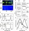
Figure 1.
Figure 1.
Phenotype, Chlorophyll Fluorescence, and Absorbance Spectra of apo1 Mutant Plants. (A) Three-week-old mutant plants growing on sucrose medium at 10 μmol photons m−2 s−1 are pale and show a hcf phenotype under UV light. WT, Wild type. (B) Chlorophyll fluorescence induction and qP at low (3 μmol photons m−2 s−1) and moderate (30 μmol photons m−2 s−1) light intensities. Leaves were exposed to a series of superimposed 800-ms saturating light flashes. (C) Absorbance spectra of mutant and wild-type leaves grown at 10 μmol photons m−2 s−1. The increased band at 473 nm (see arrows) in the second order derivative spectra (dotted line) is indicative of an increased carotenoid accumulation in the mutant relative to chlorophyll levels. (D) Low-temperature (77K) fluorescence emission spectra. The PSII band at 688 nm in the apo1 mutant was normalized to the PSI band at 735 nm in the wild type.
Figure 2.
Figure 2.
Accumulation of Plastid Proteins. Loading 100 corresponds to 8 μg of membrane or soluble proteins of the wild type. The quantity of apo1 thylakoid membranes (100%) was adjusted to wild-type levels of the ATP synthase. For quantification, dilution series of the wild type were used. Three-week-old wild-type and apo1 leaves were used for analysis. (A) Immunoblot analysis of thylakoid membrane and soluble plastid proteins. Designations of proteins and of the corresponding complexes are labeled at left and right, respectively. (B) Immunoblot analysis of the outer PSI and PSII antenna proteins in apo1 and wild type. Lhc, Light-harvesting complex. (C) Immunoblot analysis of Fe-S cluster–containing plastid proteins. The plastid-encoded thylakoid membrane and the nuclear-encoded soluble protein complexes NDH and FTR, respectively, contain [4Fe-4S] clusters. Ferredoxin (FD) binds [2Fe-2S] clusters.
Figure 3.
Figure 3.
Quantities and Integrity of psaA-psaB Transcripts and Protein Labeling Studies. (A) RNA gel blot analysis of the plastid PSI genes and the nuclear cab and rbcS genes. Eight and two (1/4) μg of total RNA from three-week-old mutant and wild-type leaves were analyzed using gene-specific probes. Sizes of the standard (right) and the bands (left) are indicated in kilobases. (B) Primer extension analysis of mutant and wild-type mRNA shows that the transcript 5′ termini at position −199 nt relative to the start codon of the psaA message are intact in apo1. (C) In vivo labeling of plastid soluble (S) and membrane (M) proteins separated by SDS-PAGE in apo1, hcf145, and the wild type (WT). Wild-type and mutant proteins with equivalent amounts of radioactivity (100.000 cpm) were loaded. (D) Polysome sedimentation in 15% to 55% sucrose (Suc) gradients by ultracentrifugation and subsequent RNA gel blot analysis of fractionated samples. Probes used are indicated at left. The filter used for the psaC probe was rehybridized with the psaA-psaB probe. The filter of the lincomycin-treated (LM) material was used for psaA-psaB and rehybridized with the psbA probe. Lincomycin treatment of wild-type and mutant plants was performed 4 h before polysome preparation. rRNAs have been detected by staining the blots.
Figure 4.
Figure 4.
Chloroplast Ultrastructure of the Wild Type and the PSI Mutants apo1, hcf101, hcf140, and hcf113. Bars = 1 μm.
Figure 5.
Figure 5.
Inactivation of APO1 by T-DNA Insertion and Complementation of apo1. (A) Schematic view of the T-DNA insertion in APO1. The two introns are indicated by shaded boxes. HindIII (H) and EcoRI (E) restriction sites and the T-DNA left (LB) and right (RB) borders are shown. Sizes are given in kilobases. The transcription start and stop are indicated. The arrows show the positions of primers used in (C). The position of the translational start (ATG) and stop (TAA) codons are indicated. (B) Genomic DNA gel blot analysis of EcoRI and HindIII double-digested mutant and wild-type (WT) DNA results in polymorphisms that can be deduced from (A). The used probes of the T-DNA right border (RB) and the APO1 gene recognize one and the same 2.8-kb fragment. Het, Heterozygous plants for apo1. (C) PCR analysis of wild-type, mutant, complemented mutant lines (apo1c), and the apo1 cDNA. Control primers, which amplify another chromosomal region of 1.335 kb, were used in the same reaction with the APO1 gene-specific primers. APO1 exon-specific primers apo1-f and apo1-r2 did not amplify a 0.547-kb product of genomic DNA in the apo1 mutant and complemented lines, but a 0.349-kb fragment originating from the expressed cDNA. (D) RNA gel blot analysis of mutant and wild type (WT) was performed with a probe specific for apo1. Equal loading (8 μg) is shown by hybridization with a probe specific for 18S rDNA.
Figure 6.
Figure 6.
Sublocalization of APO1 within the Chloroplast. (A) Import of radiolabeled APO1 proteins into isolated chloroplasts and subsequent detection of gel-separated proteins by phosphor imaging. The precursor of 49 kD (P) was imported and proteolytically processed to 42 kD at the indicated periods of chloroplast incubation. After import chloroplasts were either treated (+) or not treated (−) with thermolysin to digest nonimported proteins. (B) The APO1 protein was fused to GFP (APO1-GFP) and transiently expressed in tobacco protoplasts. GFP fluorescence was exclusively found in spots inside the chloroplast as revealed by chlorophyll fluorescence (top). Transformed protoplasts were incubated with DAPI and visualized at higher magnification (bottom). Bars = 1 μm.
Figure 7.
Figure 7.
Sequence Analysis of the APO Gene Family in Arabidopsis and Rice. (A) Scheme of the primary APO1 structure with the conserved and repeated APO motifs 1 and 2. Conserved amino acid residues in group 1 and differences between motifs 1 and 2 present in this group are highlighted. (B) Sequence alignment of motifs 1 and 2 in APO1 to APO4 in Arabidopsis (At) and rice (Os). Amino acid residues that are conserved in all members are on gray background. Amino acid residues that are specific for each defined group in Arabidopsis and rice and different in the others are underlined. Conserved differences between the motifs 1 and 2 in all four groups are listed below the alignment.

Similar articles

Cited by

References

    1. Allen, J.F., and Pfannschmidt, T. (2000). Balancing the two photosystems: Photosynthetic electron transfer governs transcription of reaction centre genes in chloroplasts. Philos. Trans. R. Soc. Lond. B Biol. Sci. 355, 1351–1359. - PMC - PubMed
    1. Arnon, D.I. (1949). Copper enzymes in isolated chloroplasts. Polyphenoloxidase in Beta vulgaris. Plant Physiol. 24, 1–13. - PMC - PubMed
    1. Barkan, A. (1998). Approaches to investigating nuclear genes that function in chloroplast biogenesis in land plants. Methods Enzymol. 297, 38–57.
    1. Barkan, A., and Goldschmidt-Clermont, M. (2000). Participation of nuclear genes in chloroplast gene expression. Biochimie 82, 559–572. - PubMed
    1. Beinert, H., Holm, R.H., and Munck, E. (2000). Iron-sulfur clusters: Nature's modular, multipurpose structures. Science 277, 653–659. - PubMed

Publication types

MeSH terms

Associated data