

Intraepithelial CD8+ T-cell-count becomes a prognostic factor after a longer follow-up period in human colorectal carcinoma: possible association with suppression of micrometastasis
- PMID: 15494715
- PMCID: PMC2410024
- DOI: 10.1038/sj.bjc.6602201
Intraepithelial CD8+ T-cell-count becomes a prognostic factor after a longer follow-up period in human colorectal carcinoma: possible association with suppression of micrometastasis
Abstract
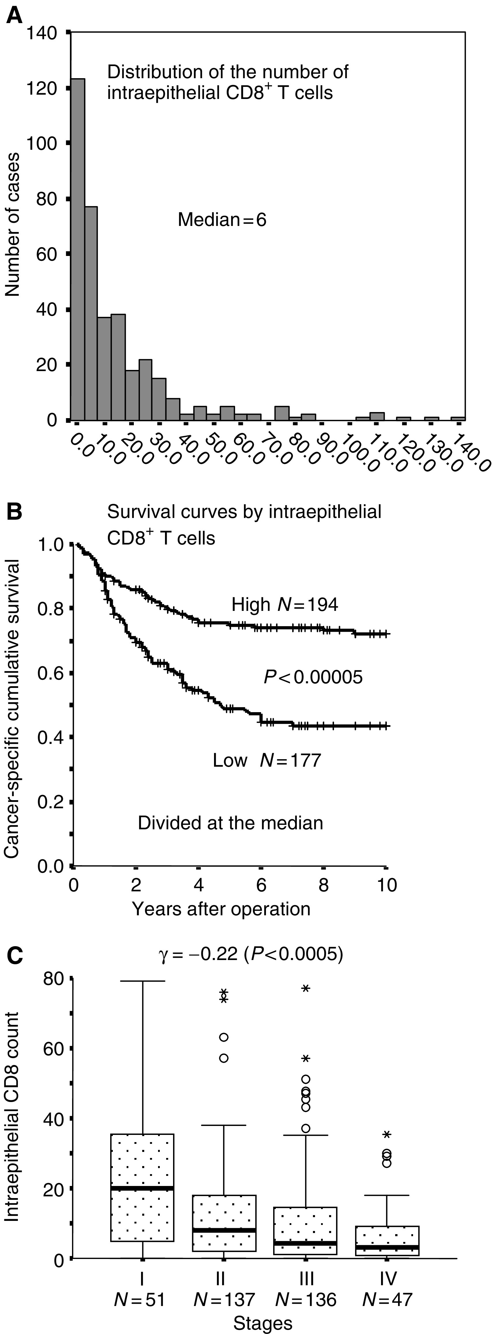
T-cell infiltration into human cancer tissues can be a manifestation of host immune responses to cancer cells. The present study was undertaken to explore the clinicopathological significance of intraepithelial CD8(+) T cells using 371 consecutively sampled human colorectal carcinomas. By univariate analysis, we noted that the survival curves by intraepithelial CD8(+) T cells became separated only after 1 to 2 years postoperation. Multivariate analyses revealed that the beneficial effect of this factor becomes significant only after a longer (more than 2 year), but not after a shorter (less than 2 year) follow-up period. Furthermore, the number of intraepithelial CD8(+) T cells was significantly higher in patients alive for more than 5 years than in patients who either died of cancer after a curative operation or patients who underwent a noncurative operation. Patients' cancer-specific death long after a curative operation is thought to be caused by the growth of micrometastases in other organs or near the primary sites. The effects of intraepithelial CD8(+) T cells, therefore, may be mediated by suppression of micrometastasis, rather than suppression of growth in the primary tumour. In conclusion, our data support a hypothesis on the presence of systemic immunosurveillance against micrometastasis of cancer cells.
Figures


References
-
- Albert ML, Darnell RB (2004) Paraneoplastic neurological degenerations: keys to tumour immunity. Nat Rev Cancer 4: 36–44 - PubMed
-
- Dolcetti R, Viel A, Doglioni C, Russo A, Guidoboni M, Capozzi E, Vecchiato N, Macri E, Fornasarig M, Boiocchi M (1999) High prevalence of activated intraepithelial cytotoxic T lymphocytes and increased neoplastic cell apoptosis in colorectal carcinomas with microsatellite instability. Am J Pathol 154: 1805–1813 - PMC - PubMed
-
- Dunn GP, Bruce AT, Ikeda H, Old LJ, Schreiber RD (2002) Cancer immunoediting: from immunosurveillance to tumor escape. Nat Immunol 3: 991–998 - PubMed
-
- Fukuzawa K, Takahashi K, Furuta K, Tagaya T, Ishikawa T, Wada K, Omoto Y, Koji T, Kakumu S (2001) Expression of fas/fas ligand (fasL) and its involvement in infiltrating lymphocytes in hepatocellular carcinoma (HCC). J Gastroenterol 36: 681–688 - PubMed
-
- Hamilton SR, Aaltonen LA (eds) (2000) WHO classification of tumours. Pathology and genetics of tumours of the digestive system. Lyon: IARC press
Publication types
MeSH terms
LinkOut - more resources
Full Text Sources
Other Literature Sources
Medical
Research Materials
