Comparative Study
. 2004 Nov 1;91(9):1669-77.
doi: 10.1038/sj.bjc.6602058.

Fhit-deficient normal and cancer cells are mitomycin C and UVC resistant

Affiliations
Comparative Study

Fhit-deficient normal and cancer cells are mitomycin C and UVC resistant

M Ottey et al. Br J Cancer. .

Abstract

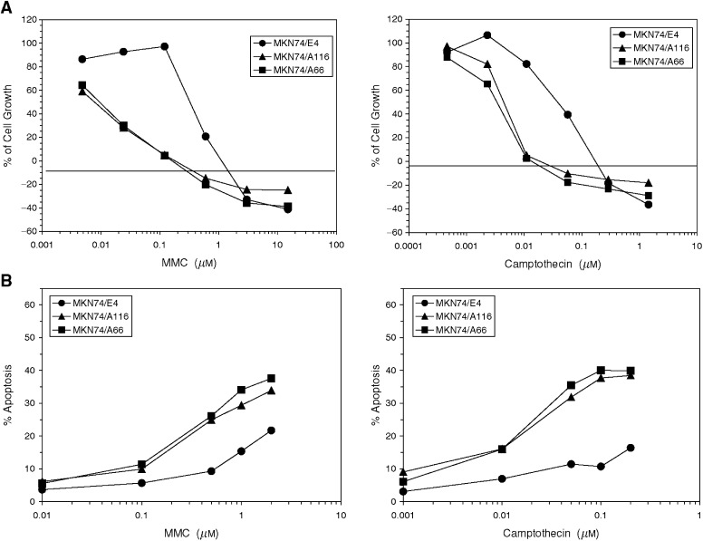
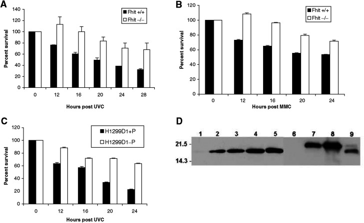
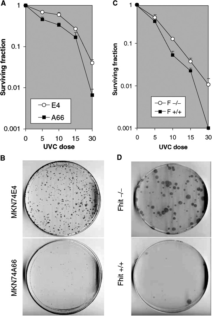
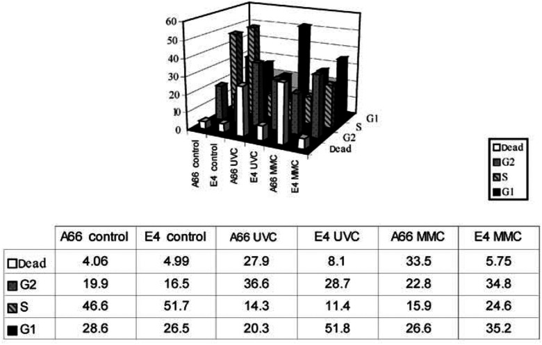
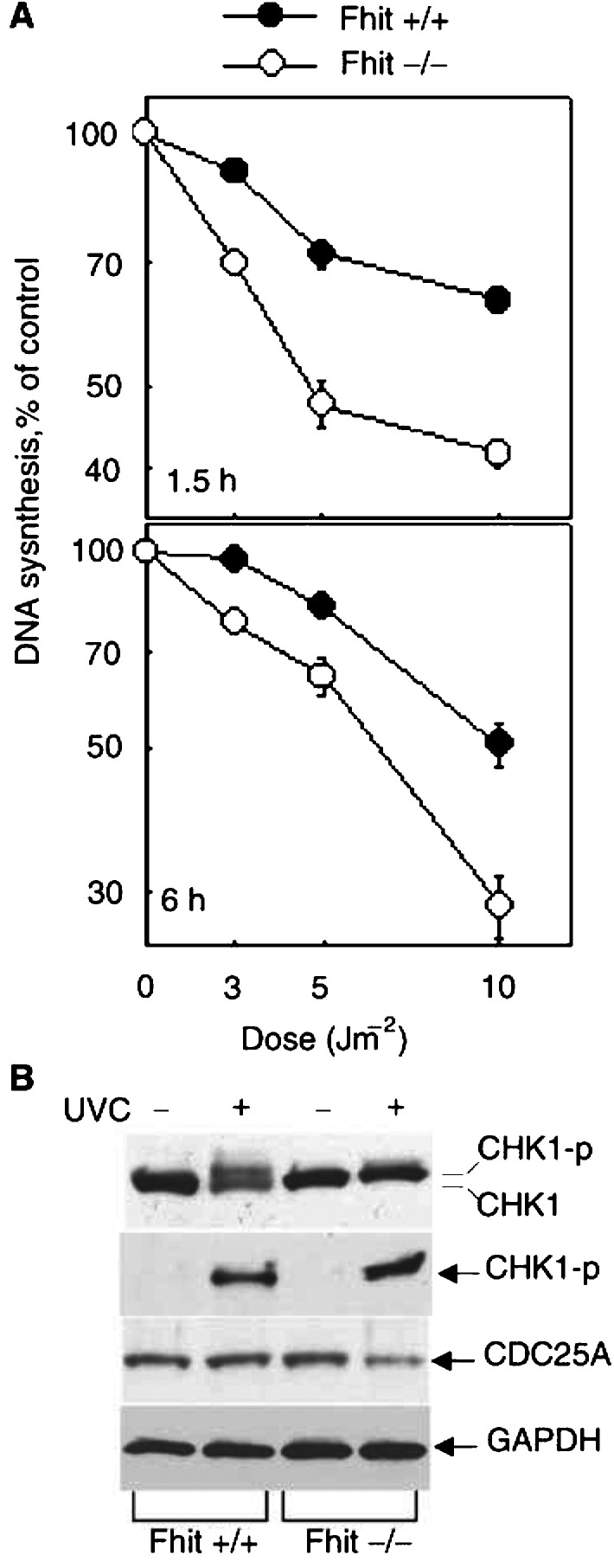
To identify functions of the fragile tumour suppressor gene, FHIT, matched pairs of Fhit-negative and -positive human cancer cell clones, and normal cell lines established from Fhit -/- and +/+ mice, were stressed and examined for differences in cell cycle kinetics and survival. A larger fraction of Fhit-negative human cancer cells and murine kidney cells survived treatment with mitomycin C or UVC light compared to matched Fhit-positive cells; approximately 10-fold more colonies of Fhit-deficient cells survived high UVC doses in clonigenic assays. The human cancer cells were synchronised in G1, released into S and treated with UVC or mitomycin C. At 18 h post mitomycin C treatment approximately 6-fold more Fhit-positive than -negative cells had died, and 18 h post UVC treatment 3.5-fold more Fhit-positive cells were dead. Similar results were obtained for the murine -/- cells. After low UVC doses, the rate of DNA synthesis in -/- cells decreased more rapidly and steeply than in +/+ cells, although the Atr-Chk1 pathway appeared intact in both cell types. UVC surviving Fhit -/- cells appear transformed and exhibit >5-fold increased mutation frequency. This increased mutation burden could explain the susceptibility of Fhit-deficient cells in vivo to malignant transformation.


Figures

Figure 1
Figure 1
Effect of genotoxic agents on cell growth and apoptosis induction in Fhit-positive (MKN74A66 and A116) and Fhit-negative (MKN74E4) gastric tumour cell lines. (A) Cell growth after 72 h exposure to mitomycin C or camptothecin, determined by MTS tetrazolium dye conversion assay. (B) Induction of apoptosis after 24 h exposure to mitomycin C or camptothecin determined by TUNEL assay.
Figure 2
Figure 2
Enhanced survival of Fhit-deficient cells after UVC and mitomycin C treatment. (A) Graphic representation of the percent of cells surviving after 60 J m−2 UVC treatment. Fhit-positive cells undergo UVC-induced apoptosis, as seen in the decreasing percent of surviving cells. Fhit-negative cells do not show a decrease in survival until 20 h after UVC treatment, and show two-fold more surviving cells by 28 h after treatment (P<0.0001, for difference between the decrease in survival of +/+ and −/− cells at 28 h). (B) Fhit +/+ and −/− cells were treated with 5 μM mitomycin C and surviving fractions determined. Fhit +/+ cells demonstrate a gradual decrease in survival over time, whereas Fhit −/− cells continue to proliferate before a more gradual decrease in survival. (Two-tailed t-test reveals that decrease in survival at 24 h is significant (P=0.0079).) Survival experiments were in duplicate or triplicate. Two-tailed Fisher's exact test computed at http://www.matforsk.no/ola/fis her.htm. (C) H1299D1+P (Fhit positive) and H1299D1-P (Fhit negative) cells were exposed to 60 J m−2 UVC. Fhit-positive cells died rapidly while Fhit-negative cells show a survival rate >60% at 24 h post UVC (P<0.0001 for survival difference at 24 h). (D) Expression of Fhit protein in Fhit-transfected cells. Lane 1: H1299D1, no induction; lanes 2–5: H1299D1 after Ponasterone A induction for 16, 24, 48, 96 h, respectively; lane 6: MKN74E4; lane 7: MKN74A66; lane 8: MKN74A116; lane 9: human lung tissue. The higher Fhit band in lanes 7 and 8 is a result of the FLAG tag (Siprashvili et al, 1997). Lane 9 shows a small amount of phosphorylated Fhit (Pekarsky et al, 2004).
Figure 3
Figure 3
Clonigenicity of UVC-treated Fhit-positive and -negative cells. (A) Dose–response clonigenicity for Fhit-positive and -negative A66 and E4 cells, respectively. MKN74E4 and A66 cells were seeded (3000 cells dish−1) after no treatment, or 5, 10, 15 and 30 J m−2 UVC. After 14 days, cells were fixed in methanol and stained with Giemsa. The values plotted are the averages from two separate experiments, containing three replica plates each. (B) MKN74E4 (Fhit negative) and MKN74A66 (Fhit positive) cells were exposed to 60 J m−2 UVC, collected, counted and re-seeded (7500 per dish). After 18 days plates were fixed in 1 : 1 methanol : acetone, stained with Giemsa and colonies counted. E4 cells formed an average of 300 colonies/7500 cells and A66 an average of 30 colonies/7500 cells. The absence of Fhit confers an advantage in surviving the lethal dose of UVC. (C) Dose–response clonigenicity for Fhit +/+ and −/− mouse cells. The cells were seeded (3000 cells dish−1) after no treatment or 5, 10, 15 or 30 J m−2 UVC. After 14 days, the cells were fixed and stained. The averages from two separate experiments, containing three replica plates each, are shown. (D) Fhit +/+ and −/− cells were exposed to 60 J m−2 UVC, collected, counted and re-seeded to dishes (5000 per dish). After 18 days, plates were fixed in 1 : 1 methanol : acetone, stained with Giemsa and colonies counted. Fhit −/− cells formed an average of 90 colonies/5000 cells and +/+ cells an average of 10 colonies/5000 cells.
Figure 4
Figure 4
Dose response of Fhit +/+ and −/− cells to UVC treatment. Fhit +/+ and −/− cells were treated with various doses of UVC, as indicated on the graph legend, collected and stained with trypan blue to determine the fraction of viable cells at each time point. At doses of 20 J m−2 or higher, Fhit +/+ cells consistently demonstrate lower viability.
Figure 5
Figure 5
UVC and mitomycin C induced cell death in Fhit-positive but not in Fhit-negative cells. Cells were synchronised first by growth in low serum and then by aphidicolin treatment for 16 h. Cells were released into S phase for 3–5 h and then left untreated (control) or mitomycin C or UVC treated. FACs analysis was performed at 18–21 h afterward, as shown for a representative experiment (21 h). The fraction of cells in specific phases of the cell cycle are illustrated in the bar graph and listed in the table below the bar graph.
Figure 6
Figure 6
(A) The rate of DNA synthesis after UVC treatment in Fhit +/+ and −/− cells. DNA synthesis was examined at 1.5 and 6 h after various doses of UVC treatment in Fhit +/+ and −/− cells. The cells pre-labelled with 14C[TdR] were changed with pre-warmed medium before UVC. At 1 and 5.5 h after exposure to different doses of UV, 0.5 μM 3H[TdR] was added to the cell culture. After 30 min, the cells were collected and loaded on GF-A filtres set in a Millipore Vacuum chamber. The rate of DNA synthesis for each sample was calculated as 3H/14C d.p.m. and is presented as a percentage of the control values obtained from untreated cells at the same time point. The data are presented as mean values and standard deviations from three independent experiments. (B) The CHK1 pathway is activated in UVC-treated Fhit-positive and -negative cells. The Fhit +/+ and −/− cells were treated with UV (15 J m−2) or untreated and harvested 2 h post treatment. Equal amounts of whole-cell extract were immunoblotted with antibodies against Chk1, phospho-Chk1 and Cdc25A, respectively. GAPDH antibody was used as the internal control for equal loading. Similar results were obtained from two independent experiments.

References

    1. Dumon KR, Ishii H, Fong LYY, Zanesi N, Fidanza V, Vecchione A, Baffa R, Trapasso F, During MJ, Huebner K, Croce CM (2001) FHIT gene therapy prevents tumor development in Fhit deficient mice. Proc Natl Acad Sci USA 98: 3346–3351 - PMC - PubMed
    1. Fong LYY, Fidanza V, Zanesi N, Lock L, Siracusa L, Mancini R, Siprashvili Z, Ottey M, Martin SE, Druck T, McCue PA, Croce CM, Huebner K (2000) Muir–Torre-like syndrome in Fhit deficient mice. Proc Natl Acad Sci USA 97: 4742–4747 - PMC - PubMed
    1. Guo Z, Kumagai A, Wang SX, Dunphy WG (2000) Requirement for Atr in phosphorylation of Chk1 and cell cycle regulation in response to DNA replication blocks and UV-damaged DNA in Xenopus egg extracts. Genes Dev 14: 2745–2756 - PMC - PubMed
    1. Heffernan TP, Simpson DA, Frank AR, Heinloth AN, Paules RS, Cordeiro-Stone M, Kaufmann WK (2002) An ATR- and Chk1-dependent S checkpoint inhibits replicon initiation following UVC-induced DNA damage. Mol Cell Biol 22: 8552–8561 - PMC - PubMed
    1. Hu B, Han SY, Wang X, Ottey M, Potoczek MB, Dicker A, Huebner K, Wang Y (2004) Involvement of the Fhit gene in the ionizing radiation-activated ATR/CHK1 pathway. J Cell Physiol, in press - PubMed

Publication types

MeSH terms