Review
. 2004 Nov;204(4):438-49.
doi: 10.1002/path.1650.

The cytoskeleton in neurodegenerative diseases

Affiliations
Review

The cytoskeleton in neurodegenerative diseases

Nigel J Cairns et al. J Pathol. 2004 Nov.

Abstract

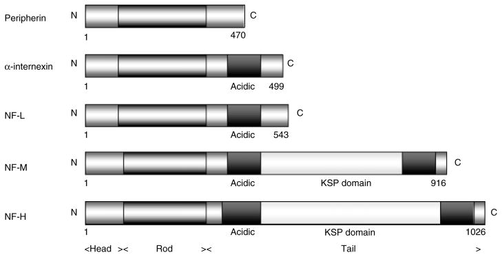
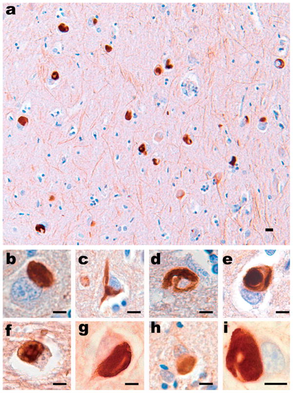
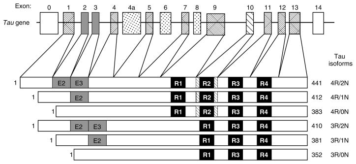
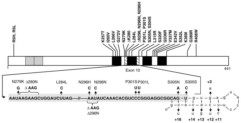
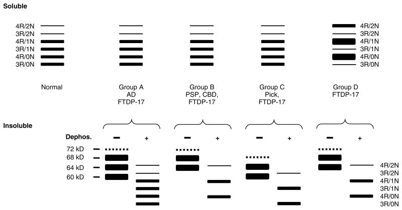
Abundant abnormal aggregates of cytoskeletal proteins are neuropathological signatures of many neurodegenerative diseases that are broadly classified by filamentous aggregates of neuronal intermediate filament (IF) proteins, or by inclusions containing the microtubule-associated protein (MAP) tau. The discovery of mutations in neuronal IF and tau genes firmly establishes the importance of neuronal IF proteins and tau in the pathogenesis of neurodegenerative diseases. Multiple IF gene mutations are pathogenic for Charcot-Marie-Tooth (CMT) disease and amyotrophic lateral sclerosis (ALS)--in addition to those in the copper/zinc superoxide dismutase-1 (SOD1) gene. Tau gene mutations are pathogenic for frontotemporal dementia with parkinsonism linked to chromosome 17 (FTDP-17), and tau polymorphisms are genetic risk factors for sporadic progressive supranuclear palsy (PSP) and corticobasal degeneration (CBD). Thus, IF and tau abnormalities are linked directly to the aetiology and pathogenesis of neurodegenerative diseases. In vitro and transgenic animal models are being used to demonstrate that different mutations impair protein function, promote tau fibrilization, or perturb tau gene splicing, leading to aberrant and distinct tau aggregates. For recognition of these disorders at neuropathological examination, immunohistochemistry is needed, and this may be combined with biochemistry and molecular genetics to properly determine the nosology of a particular case. As reviewed here, the identification of molecular genetic defects and biochemical alterations in cytoskeletal proteins of human neurodegenerative diseases has facilitated experimental studies and will promote the development of assays of molecules which inhibit abnormal neuronal IF and tau protein inclusions.


Figures

Figure 1
Figure 1
Structure of neuronal IF proteins. All proteins share a conserved structure of a head, rod with coils forming an α-helix, and tail domains containing glutamic acid-rich sequences and repeat phosphorylation motifs of lysine–serine–proline (KSP). Figures refer to the initial and terminal amino acids of each protein
Figure 2
Figure 2
All type IV neuronal IF proteins are present in the pathological inclusions of NIFID. (a) Neuronal inclusions in the subiculum of a case of NIFID contain α-internexin. α-Internexin immunohistochemistry. Neuronal inclusions in NIFID are pleomorphic. (b) Pick body-like inclusions are the most common morphological type. (c) A flame-shaped, NFT-like inclusion. (d) A filamentous serpiginous inclusion. (e) A globose NFT-like inclusion. α-Internexin immunohistochemistry. Epitopes of NF triplet proteins are present in inclusions of NIFID and are recognized by (f) phosphorylation-dependent NF-H; (g) non-phosphorylation-dependent NF-H; (h) phosphorylation-independent NF-M; and (i) phosphorylation-independent NF-L antibodies. NF immunohistochemistry. Scale bars = 10 μm
Figure 3
Figure 3
Schematic representation of the human tau gene and six human CNS tau isoforms generated by alternative splicing. The human tau gene contains 16 exons, including exon 0 that is part of the promoter. Exons 1, 4, 5, 7, 9, and 11–13 are constitutively expressed. Alternative splicing of exons 2 (E2), 3 (E3), and 10 produces the six alternative tau isoforms. Exons 6 and 8 are not transcribed in the human CNS. Exon 4a, which is also not transcribed in the human CNS, is expressed in the PNS leading to the larger tau isoforms, termed ‘big tau’. The black bars depict the 18 amino acid MT binding repeats and are designated R1 to R4. The relative sizes of the exons and introns are not drawn to scale
Figure 4
Figure 4
Schematic representation of mutations in the tau gene in FTDP-17. The structure of the largest tau isoform is shown, with known coding region mutations indicated above. The grey boxes near the amino terminus represent the alternatively spliced inserts encoded for by exons 2 and 3, while the black boxes represent each of the four MT binding repeats (not drawn to scale). The second MT binding repeat is encoded by exon 10. Part of the mRNA sequence encoding exon 10 and the intron following exon 10 is enlarged to visualize the 5′ splice site as well as the mutations both in exon 10 and within the 5′ splice site. Nucleotides that are part of intron 10 are shown in lower case
Figure 5
Figure 5
Schematic representation of western blot banding patterns of soluble and insoluble tau from different tauopathies. The drawing depicts the typical banding pattern of soluble tau (top panels) and insoluble/filamentous tau (bottom panels) from the brains of patients with FTDP-17 as well as sporadic tauopathies following resolution with SDS-PAGE and immunoblotting with anti-tau antibodies. The FTDP-17 mutations show several different western blot banding patterns of soluble and insoluble tau protein that are depicted as groups A to D. The soluble fraction from the brains of unaffected (normal) individuals, sporadic tauopathies, and FTDP-17 with mutations that do not affect tau splicing (groups A, B, and C) shows expression of all six tau isoforms. Insoluble tau from the brains of patients with FTDP-17, group A (S320F, V337M, K369I, G389R, and R406W), resolves as three major proteins of 68, 64 and 60 kD; and a minor band of 72 kD similar to that observed in AD. When dephosphorylated, they resolve into six proteins that correspond to all six tau isoforms similar to the soluble fraction. In FTDP-17 group B (R5H, P301L, and G342V), two prominent 68- and 64-kD protein bands are detected (the 72 kD minor band is variably detected) that align with 4R tau following dephosphorylation similar to that observed in PSP and CBD, indicating the selective aggregation of 4R tau. In FTDP-17 group C (K257T) and Pick’s disease, the 64 and 60 kD insoluble tau protein isoforms predominate and align with 3R tau isoforms following dephosphorylation, indicating selective aggregation of 3R tau. In contrast, in FTDP-17 mutations that affect mRNA splicing (group D: N279K, L284L, N296N, N296H, S305S, S305N, and intron 10 mutations), there is expression of predominantly 4R tau throughout the entire brain, which is reflected in the insoluble tau aggregates

Similar articles

Cited by

References

    1. Lee K, Cleveland DW. Neuronal intermediate filaments. Annu Rev Neurosci. 1996;19:187–217. - PubMed
    1. Lee VM-Y, Goedert M, Trojanowski JQ. Neurodegenerative tauopathies. Annu Rev Neurosci. 2001;24:1121–1159. - PubMed
    1. Goedert M, Spillantini MG, Serpell LC, et al. From genetics to pathology: tau and alpha-synuclein assemblies in neurodegenerative diseases. Philos Trans R Soc London Ser B Biol Sci. 2001;356:213–227. - PMC - PubMed
    1. Cleveland DW, Rothstein JD. From Charcot to Lou Gehrig: deciphering selective motor neuron death in ALS. Amyotroph Lateral Scler Other Motor Neuron Disord. 2002;3:55–56. - PubMed
    1. Al-Chalabi A, Miller CCJ. Neurofilaments and neurological disease. Bioessays. 2003;25:346–365. - PubMed

Publication types

MeSH terms