

A structural basis for the acute effects of HIV protease inhibitors on GLUT4 intrinsic activity
- PMID: 15496402
- PMCID: PMC1403823
- DOI: 10.1074/jbc.M410826200
A structural basis for the acute effects of HIV protease inhibitors on GLUT4 intrinsic activity
Abstract
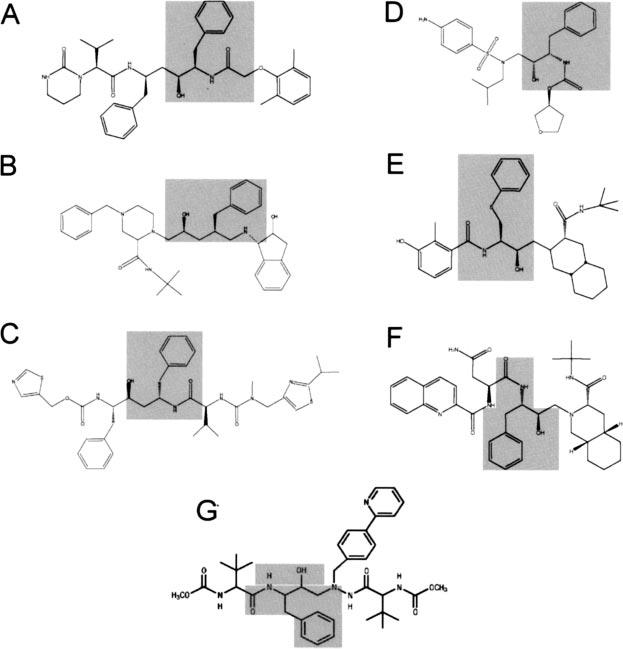
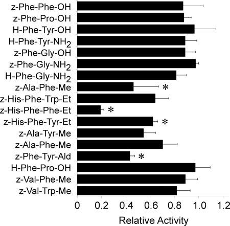
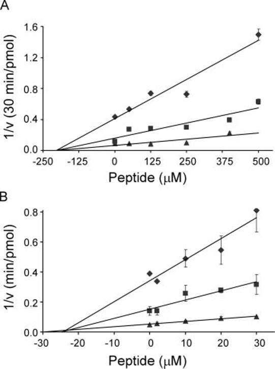
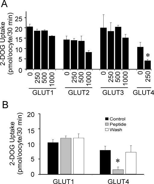
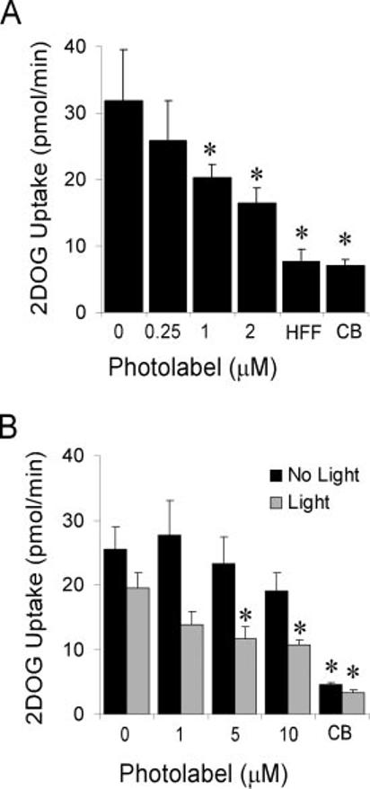
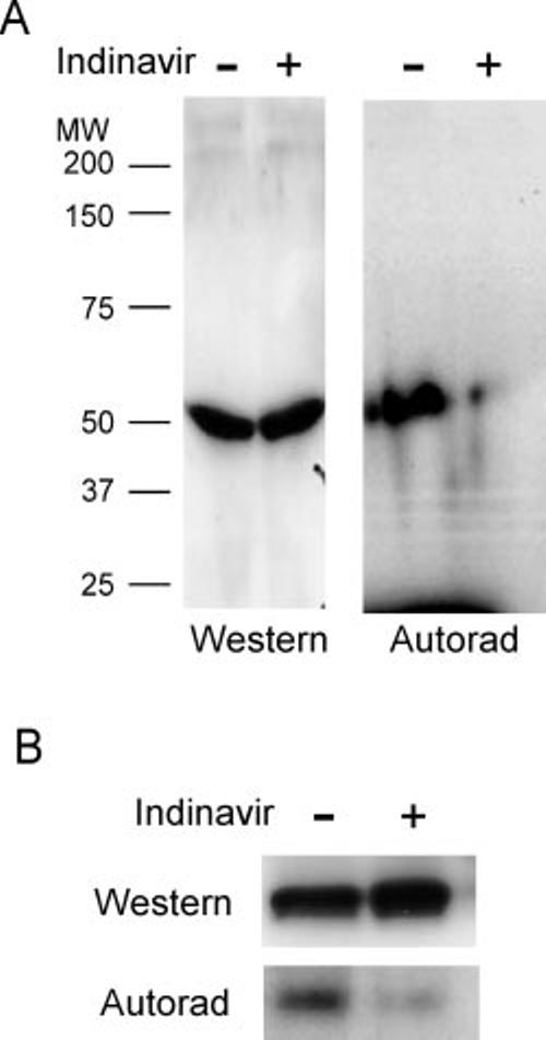
Human immunodeficiency virus (HIV) protease inhibitors (PIs) act as reversible noncompetitive inhibitors of GLUT4 with binding affinities in the low micromolar range and are known to contribute to alterations in glucose homeostasis during treatment of HIV infection. As aspartyl protease inhibitors, these compounds all possess a core peptidomimetic structure together with flanking hydrophobic moieties. To determine the molecular basis for GLUT4 inhibition, a family of related oligopeptides containing structural elements found in PIs was screened for their ability to inhibit 2-deoxyglucose transport in primary rat adipocytes. The peptide oxybenzylcarbonyl-His-Phe-Phe-O-ethyl ester (zHFFe) was identified as a potent inhibitor of zero-trans glucose flux with a K(i) of 26 mum. Similar to PIs, transport inhibition by this peptide was acute, noncompetitive, and reversible. Within a Xenopus oocyte expression system, zHFFe acutely and reversibly inhibited GLUT4-mediated glucose uptake, whereas GLUT1 activity was unaffected at concentrations as high as 1 mm. The related photoactivatable peptide zHFF-p-benzoylphenylalanine-[(125)I]Tyr-O-ethyl ester selectively labeled GLUT4 in rat adipocytes and indinavir effectively protected against photolabeling. Furthermore, GLUT4 bound to a peptide affinity column containing the zHFF sequence and was eluted by indinavir. These data establish a structural basis for PI effects on GLUT4 activity and support the direct binding of PIs to the transport protein as the mechanism for acute inhibition of insulin-stimulated glucose uptake.
Figures

Similar articles
-
The mechanism of insulin resistance caused by HIV protease inhibitor therapy.J Biol Chem. 2000 Jul 7;275(27):20251-4. doi: 10.1074/jbc.C000228200. J Biol Chem. 2000. PMID: 10806189
-
Indinavir inhibits the glucose transporter isoform Glut4 at physiologic concentrations.AIDS. 2002 Apr 12;16(6):859-63. doi: 10.1097/00002030-200204120-00005. AIDS. 2002. PMID: 11919487
-
HIV protease inhibitors act as competitive inhibitors of the cytoplasmic glucose binding site of GLUTs with differing affinities for GLUT1 and GLUT4.PLoS One. 2011;6(9):e25237. doi: 10.1371/journal.pone.0025237. Epub 2011 Sep 23. PLoS One. 2011. PMID: 21966466 Free PMC article.
-
Indinavir uncovers different contributions of GLUT4 and GLUT1 towards glucose uptake in muscle and fat cells and tissues.Diabetologia. 2003 May;46(5):649-58. doi: 10.1007/s00125-003-1080-1. Epub 2003 Apr 24. Diabetologia. 2003. PMID: 12712244
-
Intracellular organization of insulin signaling and GLUT4 translocation.Recent Prog Horm Res. 2001;56:175-93. doi: 10.1210/rp.56.1.175. Recent Prog Horm Res. 2001. PMID: 11237212 Review.
Cited by
-
HIV protease inhibitors: a review of molecular selectivity and toxicity.HIV AIDS (Auckl). 2015 Apr 8;7:95-104. doi: 10.2147/HIV.S79956. eCollection 2015. HIV AIDS (Auckl). 2015. PMID: 25897264 Free PMC article. Review.
-
The neuroenergetics of stress hormones in the hippocampus and implications for memory.Front Neurosci. 2015 May 6;9:164. doi: 10.3389/fnins.2015.00164. eCollection 2015. Front Neurosci. 2015. PMID: 25999811 Free PMC article. Review.
-
SLC2A8 (GLUT8) is a mammalian trehalose transporter required for trehalose-induced autophagy.Sci Rep. 2016 Dec 6;6:38586. doi: 10.1038/srep38586. Sci Rep. 2016. PMID: 27922102 Free PMC article.
-
Novel Roles for the Insulin-Regulated Glucose Transporter-4 in Hippocampally Dependent Memory.J Neurosci. 2016 Nov 23;36(47):11851-11864. doi: 10.1523/JNEUROSCI.1700-16.2016. J Neurosci. 2016. PMID: 27881773 Free PMC article.
-
Effects of switching from lopinavir/ritonavir to atazanavir/ritonavir on muscle glucose uptake and visceral fat in HIV-infected patients.AIDS. 2009 Jul 17;23(11):1349-57. doi: 10.1097/QAD.0b013e32832ba904. AIDS. 2009. PMID: 19474651 Free PMC article. Clinical Trial.
References
-
- Palella FJ, Jr., Delaney KM, Moorman AC, Loveless MO, Fuhrer J, Satten GA, Aschman DJ, Holmberg SD. N. Engl. J. Med. 1998;338:853–860. - PubMed
-
- Carr A, Samaras K, Burton S, Law M, Freund I, Chisholm DJ, Cooper DA. AIDS. 1998;12:F51–F58. - PubMed
-
- Mulligan K, Grunfeld C, Tai VW, Algren H, Pang M, Chernoff DN, Lo JC, Schambelan M. J. Acquired Immune Defic. Syndr. 2000;23:35–43. - PubMed
-
- Noor M, Lo JC, Mulligan K, Halvorsen R, Schwarz JM, Shambelan M, Grunfeld C. Antiviral Therapy. 2000;5:8.
-
- Murata H, Hruz PW, Mueckler M. J. Biol. Chem. 2000;275:20251–20254. - PubMed
Publication types
MeSH terms
Substances
Grants and funding
LinkOut - more resources
Full Text Sources
Other Literature Sources
Research Materials
Miscellaneous
