. 2004 Nov;131(22):5561-72.
doi: 10.1242/dev.01437. Epub 2004 Oct 20.

Eya1 and Six1 are essential for early steps of sensory neurogenesis in mammalian cranial placodes

Affiliations

Eya1 and Six1 are essential for early steps of sensory neurogenesis in mammalian cranial placodes

Dan Zou et al. Development. 2004 Nov.

Abstract

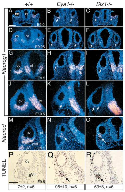
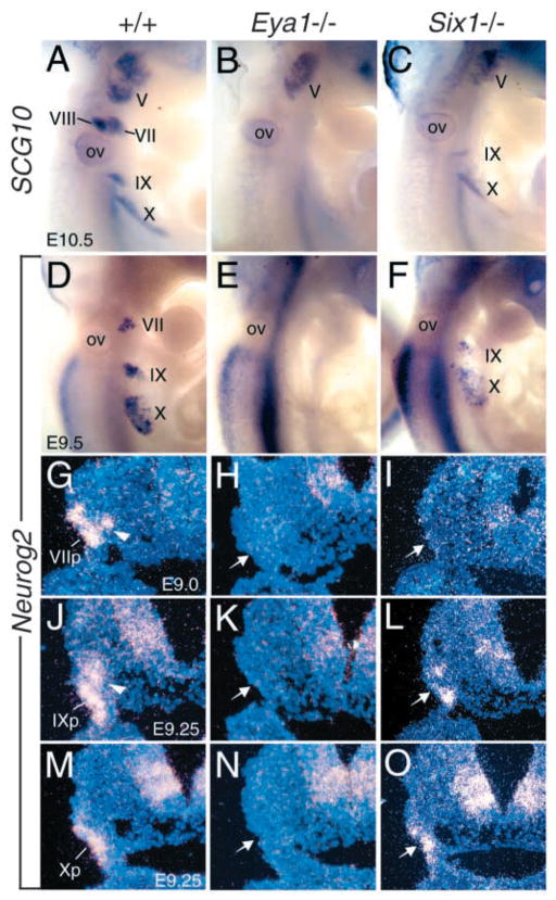
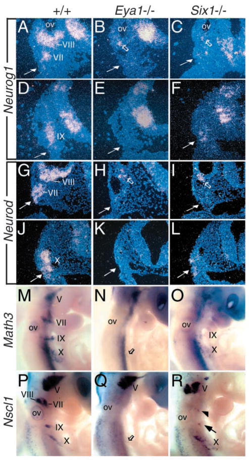
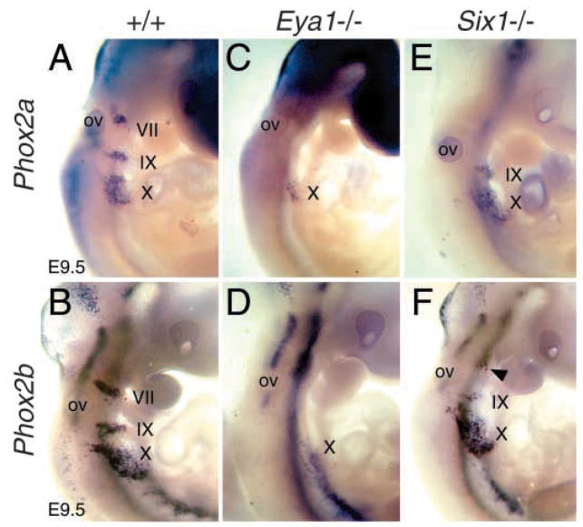
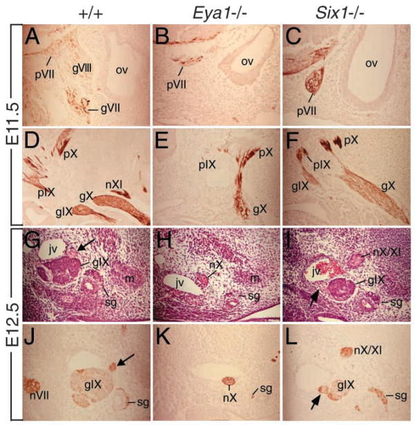
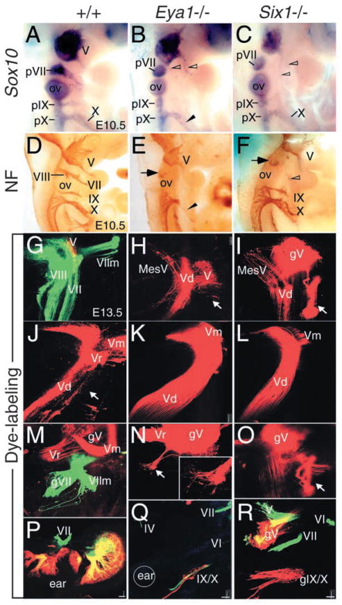
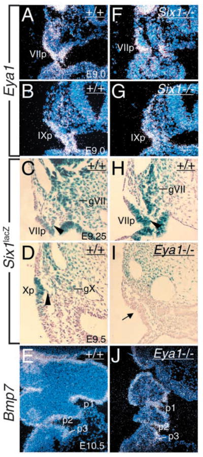
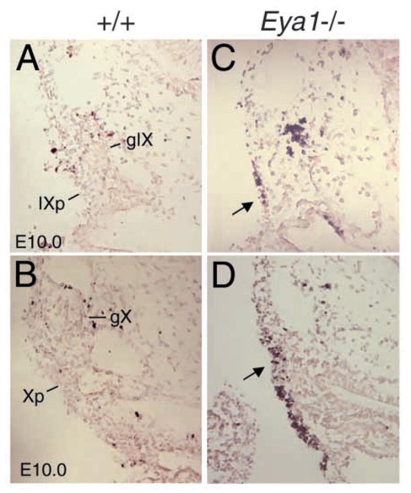
Eya1 encodes a transcriptional co-activator and is expressed in cranial sensory placodes. It interacts with and functions upstream of the homeobox gene Six1 during otic placodal development. Here, we have examined their role in cranial sensory neurogenesis. Our data show that the initial cell fate determination for the vestibuloacoustic neurons and their delamination appeared to be unaffected in the absence of Eya1 or Six1 as judged by the expression of the basic helix-loop-helix genes, Neurog1 that specifies the neuroblast cell lineage, and Neurod that controls neuronal differentiation and survival. However, both genes are necessary for normal maintenance of neurogenesis. During the development of epibranchial placode-derived distal cranial sensory ganglia, while the phenotype appears less severe in Six1 than in Eya1 mutants, an early arrest of neurogenesis was observed in the mutants. The mutant epibranchial progenitor cells fail to express Neurog2 that is required for the determination of neuronal precursors, and other basic helix-loop-helix as well as the paired homeobox Phox2 genes that are essential for neural differentiation and maintenance. Failure to activate their normal differentiation program resulted in abnormal apoptosis of the progenitor cells. Furthermore, we show that disruption of viable ganglion formation leads to pathfinding errors of branchial motoneurons. Finally, our results suggest that the Eya-Six regulatory hierarchy also operates in the epibranchial placodal development. These findings uncover an essential function for Eya1 and Six1 as critical determination factors in acquiring both neuronal fate and neuronal subtype identity from epibranchial placodal progenitors. These analyses define a specific role for both genes in early differentiation and survival of the placodally derived cranial sensory neurons.


Figures

Fig. 1
Fig. 1
Eya1 and Six1 are required for the maintenance of normal neurogenesis during inner ear morphogenesis. Section in situ hybridization on transverse sections through the otic region. (A–C) Neurog1 is expressed in the ventral otic cup (oc) of E9.0 wild-type embryos (A), and its expression in Eya1−/− (B), and Six1−/− (C) embryos is indistinguishable from wild-type embryos. (D–L) Neurog1 expression became stronger from E9.25 to 10.5 after the vesicle (ov) is closed up (D,G,J); however, its expression is markedly reduced in Eya1−/− (arrows in E,H) or Six1−/− (arrows in F,I) from E9.25, and by E10.5 only a few Neurog1-positive cells were seen in the Eya1−/− (arrow in H) or Six1−/− (arrows in L) otic vesicle. Note that Neurog1 expression in the hindbrain neural tube (nt) is relatively normal in Eya1−/− or Six1−/− embryos compared to wild-type embryos. (M–O) Neurod is expressed in the differentiating neuroblast precursors within the otic vesicle (ov) and the cells migrating to form the VIIIth ganglion (gVIII) as well as within the VIIth ganglion (gVII) from E9.5 (M) in wild-type embryos. In Eya1−/− (N) or Six1−/− (O) embryos, Neurod expression in the cells within the otic vesicle and the cells migrating away from the otic vesicle was observed (arrow in N,O). (P–R) Transverse sections through E9.5 wild-type (P), Eya1−/− (Q) and Six1−/− (R) embryos labeled with the TUNEL method for detection of apoptotic cells. TUNEL-positive cells from 6 developing VIIIth ganglia (three embryos) for each genotype were counted using an image analysis system, and the number provided represents an average number per ganglion for each genotype. Apoptosis was markedly induced in the mutants (arrow). Scale bar: 100 μm.
Fig. 2
Fig. 2
Deletion of distal cranial sensory ganglia in Eya1−/− and Six1−/− embryos. (A–C) Whole-mount hybridization with an SCG10 riboprobe of wild-type (A), Eya1−/− (B), and Six1−/− (C) embryos at E10.5. A lack of neuronal differentiation of VIIIth and distal VIIth, IXth and Xth precursors is observed in Eya1−/− embryos. In Six1−/− embryos, SCG10 staining was also missing in the regions of VIIIth and distal VIIth ganglia and slightly reduced in the regions of distal IXth and Xth ganglia. (D–F) E9.5 embryos hybridized with an Neurog2 riboprobe. Neurog2 expression in distal VII, IX and X placodes is readily visible in wild-type embryos, but its expression is completely lost in these structures in Eya1−/− embryos. In Six1−/− embryos, Neurog2 expression is undetectable in the VIIth placode and markedly reduced in the IXth and reduced in Xth placode. (G–O) Section in situ hybridization with the Neurog2 riboprobe of wild-type (G,J,M), Eya1−/− H,K,N) and Six1−/− (I,L,O) embryos at E8.5 to 9.25. Strong Neurog2 expression was observed in the distal VIIth placode (VIIp), and some migratory precursors (arrowhead) at E8.5–9.0 and in the distal IXth placode (IXp), and migratory precursors (arrowhead) at E8.75–9.25 and the Xth placode (Xp) at E9.0–9.25 in wild-type embryos, whereas its expression is completely absent in these structures of Eya1−/− at these stages (arrow, H,K,N). In Six1−/− embryos, its expression was reduced to background level in the VIIth precursors (arrow, I), also slightly reduced in the IXth (arrow, L) and relatively normal in the Xth precursors (arrow, O).
Fig. 3
Fig. 3
Alteration of Neurog1, Neurod, Math3 and Nscl1 expression in Eya1−/− and Six1−/− epibranchial neuronal precursors. (A–F) Transverse section hybridization showing Neurog1 expression in the otic vesicle (ov) and in the VIIIth ganglion region and epibranchial (VII, IX) neuronal precursors in wild-type embryos (A,D), no expression in Eya1−/− epibranchial neuronal precursors (arrow in B,E) and reduced expression in Six1−/− epibranchial neurons (arrow in C,F). Note that Neurog1 expression was observed in the VIIIth precursors (open arrow in B,C). (G–L) Transverse section hybridization showing Neurod expression in the VIIIth and epibranchial neuronal precursors in wild-type embryos (G,J), no expression in epibranchial precursors in Eya1−/− embryos (arrow in H,K) and no expression (arrow in I) or reduced expression in Six1−/− epibranchial neurons (arrow in L). Note that Neurod expression was detected in the VIIIth neuroblast precursors (open arrow in H,I). (M–O) Whole-mount hybridization showing Math3 expression in epibranchial neurons in wild-type embryos (M), only a few Math3-positive cells in Eya1−/− Xth neurons (open arrow in N), and reduced expression in Six1−/− IXth and Xth neurons (O). (P–R) Whole-mount hybridization showing Nscl1 expression in cranial sensory ganglia in wild-type embryos (P). In Eya1−/− embryos, while Nscl1 expression appeared relatively normal in the Vth ganglion, it expression is absent in VIIIth, VIIth and IXth ganglia and very few Nscl1-positive cells were detected in Eya1−/− Xth neurons (open arrow in Q). (R) Nscl1 expression is also absent in Six1−/− VIIIth ganglion and markedly reduced in Six1−/− VIIth (arrowhead) and IXth (arrow) ganglia.
Fig. 4
Fig. 4
Alteration of Phox2a and Phox2b expression in Eya1−/− and Six1−/− epibranchial neuronal precursors. Whole-mount hybridization showing Phox2a (A) and Phox2b (B) expression in the epibranchial neurons in wild-type embryos. (C,D) In Eya1−/− embryos, weak Phox2a and Phox2b expression was only observed in the Xth neurons. (E,F) In Six1−/− embryos, their expression was absent (E) or markedly reduced in the VIIth (arrowhead in F), markedly reduced in the IXth, and relatively normal in the Xth neurons.
Fig. 5
Fig. 5
Malformation of cranial sensory ganglia in Eya1−/− or Six1−/− embryos at later stages. (A–F) Neurofilament antibody staining of sagittal sections of E11.5 wild-type (A,D), Eya1−/− (B,E) and Six1−/− (C,F) heads. Neurofilament antibody labeled the proximal VIIth ganglion (pVII), which is normally fused with the VIIIth ganglion (gVIII), the proximal IXth (pIX) and Xth (pX) and the distal VIIth (gVII), IXth (gIX) and Xth (gX) ganglia in wild-type embryos (A,D). In Eya1−/− embryos, all proximal ganglia and partial structures of the distal Xth ganglion are present, but the VIIIth and the distal VIIth and IXth ganglia are missing (B,E). (C,F) In all three Six1−/− embryos, while the proximal ganglia are present, the VIIIth and distal VIIth (gVII) ganglia are missing but the distal IXth and Xth ganglia are also present. For A–F, anterior is to the left and dorsal is up. (G–I) Hematoxylin and Eosin and (J–L) neurofilament antibody staining of transverse sections of E12.5 wild-type (G,J), Eya1−/− (H,K) and Six1−/− (I,L) heads. (G,J) The IXth ganglion (gIX) and the nerve fibers near the IXth ganglion that strongly stained with both Eosin and the neurofilament antibody are present in wild-type embryos (arrow). The neurofilament antibody also strongly labeled other nerve fibers in that region, including the VIIth nerve fibers (nVII, J). (G,K) The IXth ganglion is missing in Eya1−/− embryos. However, some nerve fibers that strongly stained with both Eosin and the neurofilament antibody are present in the mutant and this structure is likely to represent the Xth nerve fibers, which are present at E11.5 (E). (I,L) The IXth ganglion is present in Six1−/− embryos with slightly smaller size and the neurofilament antibody also labeled nearby nerve fibers (arrow and nX/XI). For G–L, dorsal is up and lateral is to the left. jv, internal jugular vein; m, prevertebral premuscle mass; sg, sympathetic ganglion component.
Fig. 6
Fig. 6
Eya1 and Six1 regulate the patterning of cranial sensory nerves and their associated ganglia. (A–C) E10.5 whole-mount embryos hybridized with a Sox10 riboprobe. The proximal VIIth (pVII), IXth (pIX) and Xth (pX) ganglia are present in Eya1−/−(B) or Six1−/− (C) embryos. Truncated distal Xth ganglion (X) is present in Eya1−/− embryos (arrowhead, B). (D–F) E10.5 whole-mount embryos immunostained with an antibody against Neurofilament (NF). The VIIIth and distal VIIth, IXth and Xth ganglia are apparent in wild type embryos (D). However, the VIIIth and the distal VIIth ganglia are missing in Eya1−/− (E) and Six1−/− (F) embryos. While the distal IXth is completely absent but the distal Xth ganglion is truncated in Eya1−/− (arrowhead, E), both are present in Six1−/− embryos (F). Arrow points to the proximal VIIth ganglion in the mutants (E,F). Nerves originating from the Vth ganglion sometime innervate the second branchial arches in both mutants (open arrowheads). (G–R) Whole-mount images of E13.5 wild-type (G,J,M,P), Eya1−/− (H,K,N,Q) and Six1−/− (I,L,O,R) embryos show the development of cranial nerves using lipophilic dyes to trace the fibers from the nerves into the brain (G–O) and from the brain to the periphery (P–R). (G) An image shows the VIII projection (left two layers; vestibular and cochlear projection), the V/VII projection (green), the VII motor root (VIIm, green) and partial V projection (red) in normal animals. (H) There is no VIII nerve projection in any E13.5 Eya1−/− or (I) Six1−/− animals after applications to the hyoid (VII nerve) arch. However, the VII motoneurons were filled from the V nerve in both mutants (arrow). In addition, a V sensory component (MesV) projects not into the descending V (Vd) tract but rather into the area that receives the inner ear projection in normal animals. (J) A whole-mount image shows the V descending tract (Vd), V root (Vr) and motoneurons (Vm) in E13.5 normal animals. (K,L) Images show obvious expansion of the V projection in Eya1−/− (K) and Six1−/− (L) embryos. (M) V (red) and VII (green) ganglia and roots are shown as they approach the brain. (N) There is small projection of VII fibers out of the V ganglion in Eya1−/− animals (arrow). (O) In Six1−/− embryos, some VII motoneurons reroute directly into the V ganglion (gV) (arrow) or project outside the brain only to join the V nerve (compare images of I and O, which are taken form a lateral and a medial perspective of the same brain). (P) A motoneuron injection (green) and a sensory alar plate injection (red) caudal to the ear labels the VIII and VII nerves around the ear. (Q) In Eya1−/− embryos, only a few sensory neurons of the IX/X ganglia (red), abducens (VI), V and trochlear (IV) nerves (all green) are labeled. Circle indicates the approximate position of the ear. (R) In this Six1−/− animal, the VII motoneurons exit separately from V but join the V fibers outside the brain. Note the more profound reduction in IX/X ganglion size in Eya1 mutants (Q,R). Anterior is up and dorsal is to the left in all images. Scale bar: 100 μm.
Fig. 7
Fig. 7
Eya1 controls Six1 expression in epibranchial placodes. (A,B,F,G) Transverse sections showing Eya1 expression in distal VIIth placode (VIIp, A) at E9.0 and distal IXth placode (pIX, B) at E9.0 and its expression in these structures was preserved in Six1−/− embryos (F,G). (C,D,H,I) Transverse sections showing Six1lacZ expression in distal VIIth, IXth and Xth placodal, migratory (arrowhead) and aggregating neuronal precursors in the ganglion anlagen (gVII, gIX, gX) at E9.25–9.5 in Six1lacZ/+ heterozygous embryos (C,D,H) and its expression was undetectable in Eya1−/− epibranchial neuronal precursors (I). (E,J) Bmp7 expression in the pharyngeal pouches (p1–p3, E) at E10.5 was unaffected in Eya1−/− embryos (J).
Fig. 8
Fig. 8
Eya1−/− placodal progenitor cells undergo abnormal apoptosis. Transverse sections labeled with the TUNEL method for detection of apoptotic cells showing a few apoptotic cells in the wild-type IX (IXp, A) and X placodes (Xp, B) and more apoptotic cells in Eya1−/− IX and X placodes (arrow, C and D). gIX and gX, IXth and Xth cranial ganglia.

Similar articles

Cited by

References

    1. Abdelhak S, Kalatzis V, Heilig R, Compain S, Samson D, Vincent C, Weil D, Cruaud C, Sahly I, Leibovici M, et al. A human homologue of the Drosophila eyes absent gene underlies branchio-oto-renal (BOR) syndrome and identifies a novel gene family. Nat Genet. 1997;15:157–164. - PubMed
    1. Adam J, Myat A, Le Roux I, Eddison M, Henrique D, Ish-Horowicz D, Lewis J. Cell fate choices and the expression of Notch, Delta and Serrate homologues in the chick inner ear: parallels with Drosophila sense-organ development. Development. 1998;125:4645–4654. - PubMed
    1. Ayer-Le Lievre CS, Le Douarin NM. The early development of cranial sensory ganglia and the potentialities of their component cells studied in quail-chick chimeras. Dev Biol. 1982;94:291–310. - PubMed
    1. Begbie J, Brunet JF, Rubenstein JL, Graham A. Induction of the epibranchial placodes. Development. 1999;126:895–902. - PubMed
    1. Bonini NM, Leiserson WM, Benzer S. The eyes absent gene: genetic control of cell survival and differentiation in the developing Drosophila eye. Cell. 1993;72:379–395. - PubMed

Publication types

MeSH terms