. 2005 Jan;16(1):231-7.
doi: 10.1091/mbc.e04-07-0547. Epub 2004 Oct 20.

Palmitoylation-dependent estrogen receptor alpha membrane localization: regulation by 17beta-estradiol

Affiliations

Palmitoylation-dependent estrogen receptor alpha membrane localization: regulation by 17beta-estradiol

Filippo Acconcia et al. Mol Biol Cell. 2005 Jan.

Abstract

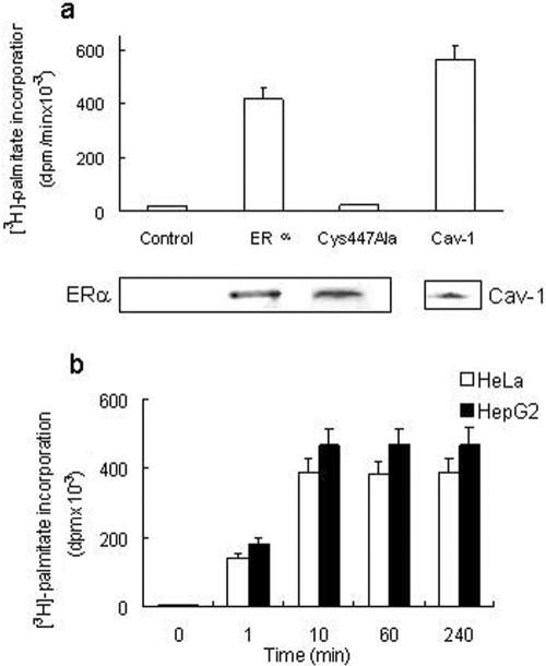
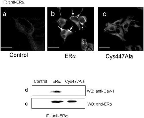
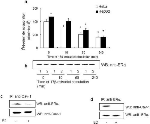
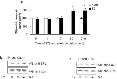
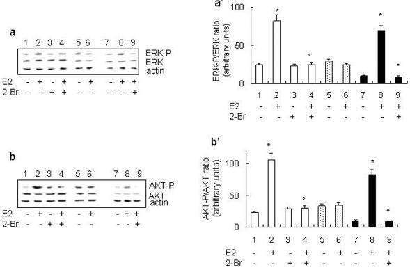
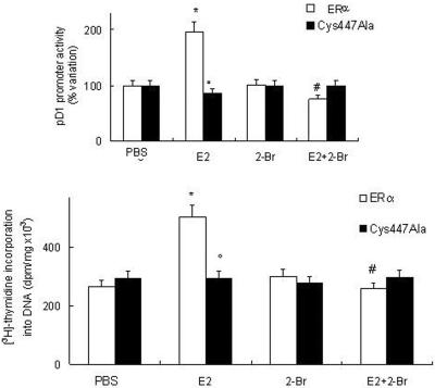
A fraction of the nuclear estrogen receptor alpha (ERalpha) is localized to the plasma membrane region of 17beta-estradiol (E2) target cells. We previously reported that ERalpha is a palmitoylated protein. To gain insight into the molecular mechanism of ERalpha residence at the plasma membrane, we tested both the role of palmitoylation and the impact of E2 stimulation on ERalpha membrane localization. The cancer cell lines expressing transfected or endogenous human ERalpha (HeLa and HepG2, respectively) or the ERalpha nonpalmitoylable Cys447Ala mutant transfected in HeLa cells were used as experimental models. We found that palmitoylation of ERalpha enacts ERalpha association with the plasma membrane, interaction with the membrane protein caveolin-1, and nongenomic activities, including activation of signaling pathways and cell proliferation (i.e., ERK and AKT activation, cyclin D1 promoter activity, DNA synthesis). Moreover, E2 reduces both ERalpha palmitoylation and its interaction with caveolin-1, in a time- and dose-dependent manner. These data point to the physiological role of ERalpha palmitoylation in the receptor localization to the cell membrane and in the regulation of the E2-induced cell proliferation.


Figures

Figure 1.
Figure 1.
ERα palmityolation. (a) [3H]Palmitate incorporation into the empty vector (control) or wild-type ERα or ERα-Cys447Ala mutant-transfected HeLa cells and into immunoprecipitated caveolin-1 (Cav-1) of untransfected HeLa cells has been evaluated after 4 h of palmitate labeling (top). Western blot analysis of the corresponding 67-kDa band of immunoprecipitated ERα or the 22-kDa band of immunoprecipitated caveolin-1 (bottom). (b) Time course of [3H]palmitate incorporation into immunoprecipitated ERα in ERα-transfected HeLa and in HepG2 cells. Data are the means of four independent experiments ± SD.
Figure 2.
Figure 2.
Subcellular localization of ERα and ERα-Cys447Ala mutant and ERα:caveolin-1 interaction. Confocal microscopy analysis of empty vector (a; control), wild-type ERα (b), and ERα-Cys447Ala mutant (c)-transfected HeLa cells by using anti-ERα MC20 antibody on freeze-thawed HeLa cells. White arrows indicate the membrane localization of wild-type ERα. In a, fluorescence signal is superimposed to laser pseudophase contrast images of the cells. Bar, 10 μm. Empty vector (control) or wild-type ERα or ERα-Cys447Ala mutant-transfected HeLa cells were subjected to ERα immunoprecipitation with anti-ERα MC20 antibody followed by Western blot with anti-caveolin-1 antibody (d) or by Western blot with anti-ERα MC20 antibody (e).
Figure 3.
Figure 3.
E2 effect on ERα palmityolation and ERα association with caveolin-1. Time course (a) and the corresponding Western blot analysis (b) of E2-stimulated ERα-transfected HeLa (open bars; 1) and HepG2 (filled bars; 2) cells. [3H]palmitate incorporation in immunoprecipitated ERα was performed at 4 h. Data are the means of four independent experiments ± SD. *p < 0.001, compared with unstimulated samples (0) were determined by using Student's t test. Wild-type ERα-transfected HeLa cells were subjected to caveolin-1 immunoprecipitation (c) or ERα immunoprecipitation (d) with anti-caveolin-1 or anti-ERα MC20 antibodies followed by Western blot with anti-caveolin-1 or with anti-ERα MC20 antibodies.
Figure 4.
Figure 4.
E2 effect on A/B and C domains deleted ERα mutant (HE14) palmityolation and association with caveolin-1. (a) Time course of E2-stimulated ERα-HE14 mutant-transfected HeLa cells. [3H]Palmitate incorporation in immunoprecipitated ERα-HE14 mutant was performed at 4 h in the absence (open bars) or presence (filled bars) of 10 nM E2. Data are the means of four independent experiments ± SD. *p < 0.001, compared with unstimulated samples (open bars), were determined by using Student's t test. ERα-HE14 mutant-transfected HeLa cells were subjected to caveolin-1 immunoprecipitation (b) or ERα immunoprecipitation (c) with anti-caveolin-1 or anti-ERα MC20 antibodies followed by Western blot with anti-caveolin-1 or with anti-ERα MC20 antibodies.
Figure 5.
Figure 5.
Effect of ERα palmitoylation on E2-induced ERK and AKT phosphorylation. Western blot analysis of ERK phosphorylation in ERα (lanes 1-4) or ERα-Cys447Ala mutant (lanes 5 and 6)-transfected HeLa and HepG2 (lanes 7-9) cells were performed on unstimulated (-) and stimulated (+) cells for 10 min with E2 (10 nM) in the presence or absence of 10 μM PAT inhibitor 2-Br (30-min pretreatment). The same filter was reprobed with anti-total ERK and anti-actin antibodies. (a) Typical Western blot. (a′) Densitometric analysis of four different experiments. Data are the means ± SD. p < 0.001, compared with unstimulated samples (*) or with E2-stimulated samples (°) were determined by using Student's t test. Western blot analysis of AKT phosphorylation in ERα (lanes 1-4) or ERα-Cys447Ala mutant (lanes 5 and 6)-transfected HeLa and HepG2 (lanes 7-9) cells were performed on unstimulated (-) and stimulated (+) cells for 30 min with E2 (10 nM) in the presence or absence of 10 μM PAT inhibitor 2-Br (30-min pretreatment). The same filter was reprobed with anti-total AKT and anti-actin antibodies. (b) Typical Western blot. (b′) Densitometric analysis of four different experiments. Data are the means ± SD. p < 0.001, compared with unstimulated samples (*) or with E2-stimulated samples (°) were determined by using Student's t test.
Figure 6.
Figure 6.
Effect of ERα palmitoylation on E2-induced cyclin D1 promoter activity and DNA synthesis. (a) Luciferase assay detection on HeLa cells cotransfected with pXP2-D1-2966-luciferase and wild-type ERα or ERα-Cys447Ala mutant expression vectors. After transfection, cells were treated with vehicle (PBS) or E2 (10 nM) in the presence or absence of 10 μM PAT inhibitor 2-Br (30-min pretreatment). (b) [3H]Thymidine incorporation into DNA in ERα or ERα-Cys447Ala mutant-transfected HeLa cells treated with vehicle (PBS) or E2 (10 nM) in the presence or in absence of 10 μM PAT inhibitor 2-Br (30-min pretreatment). Data are the means ± SD of four independent experiments. *p < 0.001, compared with respective control values (PBS); °p < 0.001, compared with ERα-transfected cells stimulated with E2 values; and #p < 0.001, compared with respective E2 values, were determined by using Student's t test.

Similar articles

Cited by

References

    1. Acconcia, F., Ascenzi, P., Fabozzi, G., Visca, P., and Marino, M. (2004b). S-Palmitoylation modulates human estrogen receptor-α functions. Biochem. Biophys. Res. Commun. 316, 878-883. - PubMed
    1. Acconcia, F., Totta, P., Ogawa, S., Cardillo, I., Inoue, S., Leone, S., Trentalance, A., Muramatsu, M., and Marino, M. (2004a). Survival versus apoptotic 17β-estradiol effect: role of ERα and ERβ activated non-genomic signalling. J. Cell. Physiol. Published online at www.interscience.wiley.com/DOI10.1002/5CP.20219. - PubMed
    1. Arvanitis, D. N., Wang, H., Bagshaw, R. D., Callahan, J. W., and Boggs, J. M. (2004). Membrane-associated estrogen receptor and caveolin-1 are present in central nervous system myelin and oligodendrocyte plasma membranes. J. Neurosci. Res. 75, 603-613. - PubMed
    1. Bijlmakers, M. J., and Marsh, M. (2003). The on-off story of protein palmitoylation. Trends Cell Biol. 13, 32-42. - PubMed
    1. Bradford, M. M. (1976). A rapid and sensitive method for the quantitation of microgram quantities of protein utilizing the principle of protein-dye binding. Anal. Biochem. 772, 248-254. - PubMed

Publication types

MeSH terms