. 2005 Jan;16(1):339-47.
doi: 10.1091/mbc.e04-05-0438. Epub 2004 Oct 20.

NGF controls dendrite development in hippocampal neurons by binding to p75NTR and modulating the cellular targets of Notch

Affiliations

NGF controls dendrite development in hippocampal neurons by binding to p75NTR and modulating the cellular targets of Notch

Patricia Salama-Cohen et al. Mol Biol Cell. 2005 Jan.

Abstract

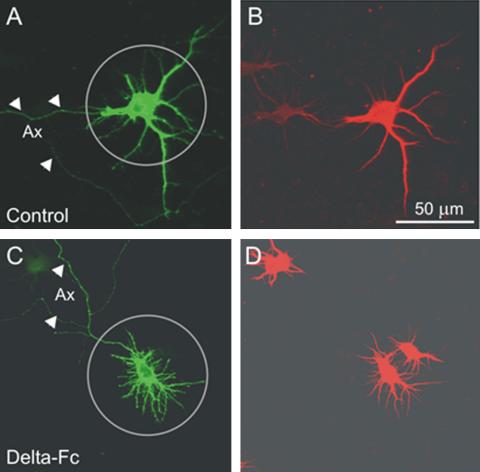
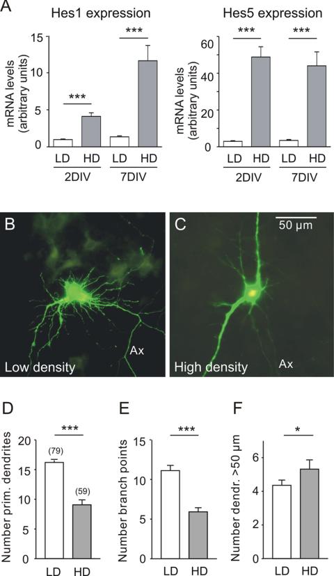
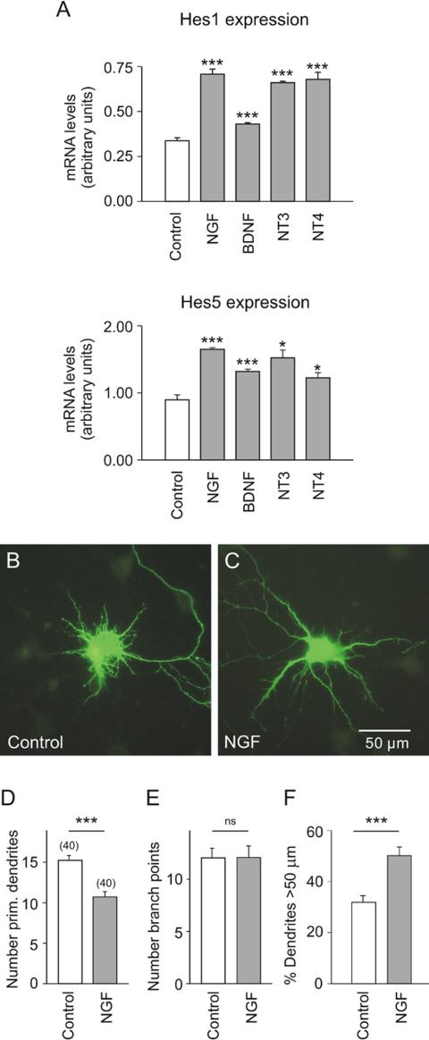
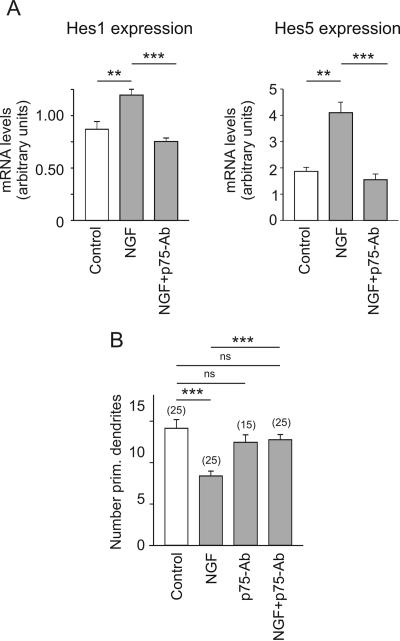
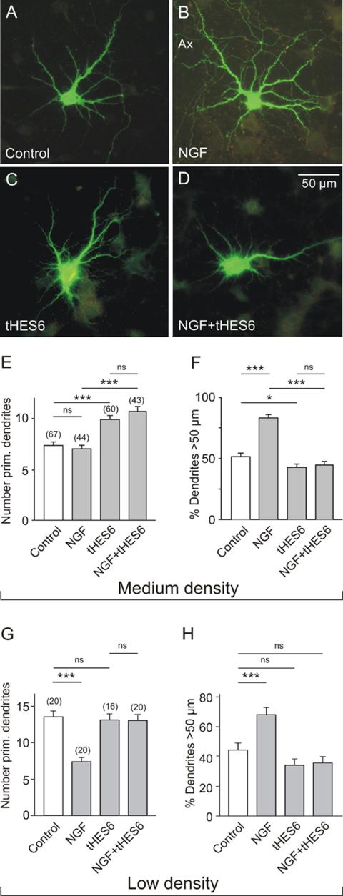
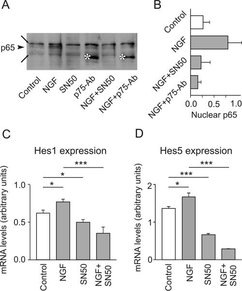
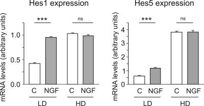
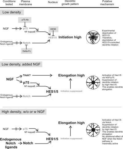
Notch and neurotrophins control neuronal shape, but it is not known whether their signaling pathways intersect. Here we report results from hippocampal neuronal cultures that are in support of this possibility. We found that low cell density or blockade of Notch signaling by a soluble Delta-Fc ligand decreased the mRNA levels of the nuclear targets of Notch, the homologues of enhancer-of-split 1 and 5 (Hes1/5). This effect was associated with enhanced sprouting of new dendrites or dendrite branches. In contrast, high cell density or exposure of low-density cultures to NGF increased the Hes1/5 mRNA, reduced the number of primary dendrites and promoted dendrite elongation. The NGF effects on both Hes1/5 expression and dendrite morphology were prevented by p75-antibody (a p75NTR-blocking antibody) or transfection with enhancer-of-split 6 (Hes6), a condition known to suppress Hes activity. Nuclear translocation of NF-kappaB was identified as a link between p75NTR and Hes1/5 because it was required for the up-regulation of these two genes. The convergence of the Notch and p75NTR signaling pathways at the level of Hes1/5 illuminates an unexpected mechanism through which a diffusible factor (NGF) could regulate dendrite growth when cell-cell interaction via Notch is not in action.


Figures

Figure 1.
Figure 1.
Identification of axons and dendrites. Shown are confocal micrographs of two neurons, one grown under control conditions (A and B) and another neuron treated with Delta-Fc, a condition that increases the number of dendrites (C and D; see Figure 3C). (A and C) Immunostaining of EGFP; (B and D) immunostaining of MAP2a/b. Note the absence of MAP-2 immunoreactivity from the longest neuron process, the putative axon (arrowheads in A and C) and the presence of MAP-2 staining on thin first- and second-order nonaxonal branches. Circles, circular region of interest (ROI), as used for morphometric evaluation of dendrites.
Figure 2.
Figure 2.
The effect of cell density on Hes1/5 mRNA levels and on dendrite morphology of EGFP-labeled hippocampal neurons in culture. (A) Neurons from E17 mouse hippocampi were plated at an initial cell density of 15,000/cm2 (LD) or 150,000/cm2 (HD) and maintained for either 2 or 7 DIV. The mRNA levels of Hes1 and Hes5 were determined by reverse transcription and quantitative PCR as described in the experimental section. Shown is that at high cell density the mRNA levels of both Hes1 and Hes5 were higher. (B and C) Digital fluorescence images of EGFP-expressing hippocampal neurons after 7 DIV. Ax, axon. Note that low cell density favored the appearance of additional first-order (primary) dendrites and second-order (secondary) dendrite branches, whereas high cell density favored dendrite elongation. (D-F) Results of morphometric evaluation. The graphs show the mean, SE, and number of observations (in brackets) for each treatment. Here and in the following, significance levels (asterisks) were determined for the data sets connected by horizontal lines using an unpaired t test. *p < 0.05, **p < 0.01, ***p < 0.001. (D) Number of primary dendrites, i.e., dendrites emerging from the soma. (E) Number of branch points. This parameter represents the total number of bifurcations of the labeled neuron within a radius of 50 μm. (F) Number of dendrite arbors that exceeded the limits of the ROI, i.e., a length >50 μm.
Figure 3.
Figure 3.
The effect of Notch blockade on Hes1/5 mRNA levels and on dendrite morphology of EGFP-labeled hippocampal neurons in culture. (A) Neurons from E17 mouse hippocampi were plated at an initial cell density of 50,000/cm2. After 7 DIV the recombinant Notch ligand Delta-Fc (14 nM) was added for 3.5 h. The mRNA levels of Hes1 and Hes5 were determined by RT and quantitative PCR. Observe that under the given experimental conditions Delta-Fc behaved as an antagonist of Notch activity, inducing a decrease in the mRNA levels of both Hes1 and Hes5. (B and C) Digital fluorescence images of EGFP-labeled hippocampal neurons. Same cell density as in A. Note Delta-Fc induced the appearance of additional primary dendrites and second-order dendrite branches and decreased the fraction of dendrites >50 μm. (D-F) Summary of morphometric evaluation. The difference in absolute number of dendrites >50 μm was not significantly different in this experiment. Symbols and parameters as in Figure 2.
Figure 4.
Figure 4.
The effect of neurotrophins/NGF on Hes1/5 mRNA levels and on dendrite morphology of EGFP-labeled hippocampal neurons in culture. (A) Results of quantitative RT-PCR experiments on hippocampal neurons that were plated at an initial cell density of 30,000/cm2 and after 7 DIV for 3.5 h exposed to the corresponding neurotrophin at a final concentration of 100 ng/ml. Shown is that all neurotrophins induced an increase of Hes1 and Hes5 mRNA. (B-F) Data from low-density cultures (15,000/cm2). Transfection on DIV 7 and exposure to NFG (100 ng/ml) for 16 h. Observe that in the controls the number of primary dendrites was higher (B and D). The presence of NGF decreased the number of primary dendrites and promoted dendrite elongation (C and F). Symbols and parameters as in Figure 2.
Figure 5.
Figure 5.
The effect of p75NTR blockade on Hes1/5 mRNA levels and on dendrite morphology of EGFP-labeled hippocampal neurons in culture. (A) Results of quantitative RT-PCR experiments on hippocampal neurons that were plated at an initial density of 15,000/cm2 and after 7 DIV for 3.5 h exposed to standard medium (control), NGF (100 ng/ml) or NGF plus blocking p75-antibody (1:100). Observe that the presence of the antibody prohibited the NGF-induced increase in the mRNA levels of Hes1 and Hes5. (B) Results of morphometric evaluation in cultures of 15,000 cells/cm2. Observe that the presence of the p75NTR-blocking antibody prevented the NGF-induced decrease in the number of primary dendrites. Symbols and parameters as in Figure 2.
Figure 6.
Figure 6.
Effect of Hes6 expression on the dendrite morphology of hippocampal neurons in medium- and low-density cultures. (A-D) Digital fluorescence images of EGFP-labeled neurons in medium-density cultures after 16 h in control medium (A and C) or in medium with NGF (100 ng/ml; B and D). E17 hippocampal neurons were cultured for 7 DIV and then transfected with pIRES vectors that either induced expression of EGFP alone (A and B) or the expression of EGFP and Hes6 (C and D). Note the larger dendritic field of the NGF-treated neuron (B) and the lack of response to NGF in the cell expressing Hes6 (D). (E and F) Results of morphometric evaluation of medium density cultures (50,000 cells/cm2). Note that NGF was unable to prevent the Hes6-stimulated outgrowth of new primary dendrites. Hes6-transfection also effectively blocked the NGF-induced dendrite elongation. (G and H) Results of morphometric evaluation of low cell density cultures (15,000 cells/cm2). Note that NGF decreased the number of primary dendrites and increased the relative length of dendrites, but these effects were prevented by transfection with Hes6. Symbols and parameters as in Figure 2.
Figure 7.
Figure 7.
NGF promotes the migration of p65/RelA to the nucleus, a process necessary for the increase of Hes1 and Hes5 mRNA. (A) Hippocampal neurons were plated at an initial density of 30,000/cm2. After 7 DIV they were for 3.5 h incubated in NGF, SN50 (an inhibitor of NF-kappaB migration into the nucleus, used here at 28 μg/ml), p75-antibody, NGF plus SN50, or NGF plus p75-antibody (control, no treatment) and then processed to obtain nuclear extracts. The same amount of nuclear protein was separated by SDS/PAGE and transferred to nitrocellulose membranes, which were subsequently probed with an anti-p65 antibody. Note the presence of p65 in the nucleus after treatment with NGF (arrowhead). Both SN50 and p75-antibody prevented the NGF-induced appearance of p65 in the nucleus. Long arrows point to nonspecific bands, asterisks to the heavy chain of the p75-antibody. (B) The intensity of the p65 band was determined by densitometry. Shown is the quantitative analysis from three different experiments. (C) Cultured hippocampal neurons were treated with NGF, with NGF plus SN50, or with SN50 alone. The presence of the inhibitor of the nuclear translocation of NF-kappaB prevented the NGF-induced increase of both Hes1 and Hes5 mRNA.
Figure 8.
Figure 8.
Quantitative PCR experiments to show the cell-density-dependent effect of NGF on Hes1/5 mRNA levels. Hippocampal neurons were for 7 DIV cultured at low (LD, 15,000 cells/cm2) or high (HD, 150,000 cells/cm2) cell density and for 3.5 h incubated in the absence or presence of NGF (100 ng/ml). Observe that NGF increased Hes1/5 mRNA levels only in low cell density cultures.
Figure 9.
Figure 9.
Summarizing scheme to illustrate experimental manipulations (shaded fields), molecular interactions and resultant effects on dendrite growth.

Similar articles

Cited by

References

    1. Aibel, L., Martin-Zanca, D., Perez, P., and Chao, M. V. (1998). Functional expression of TrkA receptors in hippocampal neurons. J. Neurosci. Res. 54, 424-431. - PubMed
    1. Bae, S., Bessho, Y., Hojo, M., and Kageyama, R. (2000). The bHLH gene Hes6, an inhibitor of Hes1, promotes neuronal differentiation. Development 127, 2933-2943. - PubMed
    1. Baker, R. E., Dijkhuizen, P. A., Van Pelt, J., and Verhaagen, J. (1998). Growth of pyramidal, but not non-pyramidal, dendrites in long-term organotypic explants of neonatal rat neocortex chronically exposed to neurotrophin-3. Eur. J. Neurosci. 10, 1037-1044. - PubMed
    1. Bash, J., Zong, W. X., Banga, S., Rivera, A., Ballard, D. W., Ron, Y., and Gelinas, C. (1999). Rel/NF-kappaB can trigger the Notch signaling pathway by inducing the expression of Jagged1, a ligand for Notch receptors. EMBO J. 18, 2803-2811. - PMC - PubMed
    1. Bentley, C. A., and Lee, K. F. (2000). p75 is important for axon growth and Schwann cell migration during development. J. Neurosci. 20, 7706-7715. - PMC - PubMed

Publication types

MeSH terms