

The Ets-1 transcription factor is required for complete pre-T cell receptor function and allelic exclusion at the T cell receptor beta locus
- PMID: 15496469
- PMCID: PMC524847
- DOI: 10.1073/pnas.0405546101
The Ets-1 transcription factor is required for complete pre-T cell receptor function and allelic exclusion at the T cell receptor beta locus
Abstract
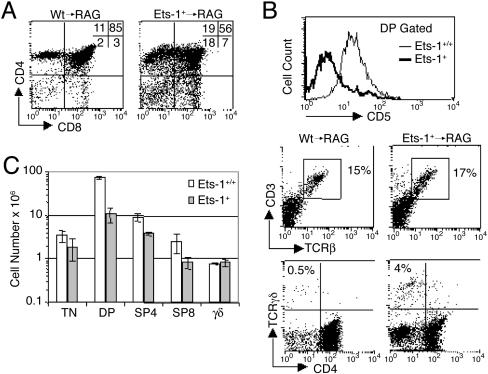
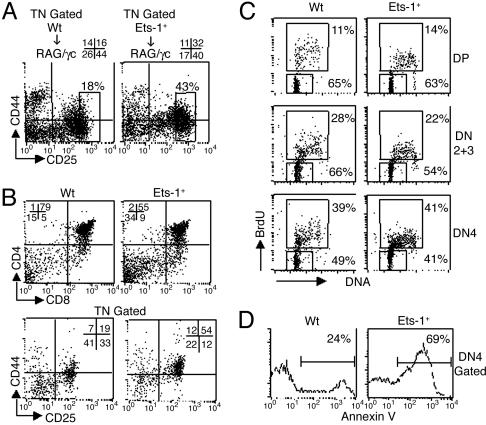
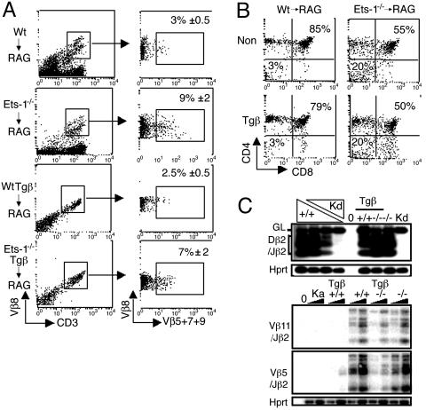
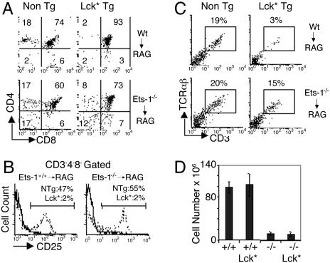
The pre-T cell receptor (TCR) functions as a critical checkpoint during alphabeta T cell development. Signaling through the pre-TCR controls the differentiation of immature CD4(-)CD8(-)CD25(+)CD44(-) [double-negative (DN)3] thymocytes into CD4(+)CD8(+) double-positive (DP) cells through the CD4(-)CD8(-)CD25(-)CD44(-)(DN4) stage. In addition, pre-TCR activity triggers expansion and survival of thymocytes and inhibits TCRbeta gene rearrangement through a process referred to as allelic exclusion. Whereas many proteins involved in the pre-TCR transduction cascade have been identified, little is known about the nuclear factors associated with receptor function. Here, we use gene targeting to inactivate the Ets-1 transcription factor in mice and analyze pre-TCR function in developing Ets-1-deficient (Ets-1(-/-)) thymocytes. We find that inactivation of Ets-1 impairs the development of DN3 into DP thymocytes and induces an elevated rate of cell death in the DN4 subset. This defect appears specific to the alphabeta lineage because gammadelta T cells maturate efficiently. Finally, the percentage of thymocytes coexpressing two different TCRbeta chains is increased in the Ets-1(-/-) background and, in contrast with wild type, forced activation of pre-TCR signaling does not block endogenous TCRbeta gene rearrangement. These data identify Ets-1 as a critical transcription factor for pre-TCR functioning and for allelic exclusion at the TCRbeta locus.
Figures
Similar articles
-
T lymphocyte development in p56lck deficient mice: allelic exclusion of the TcR beta locus is incomplete but thymocyte development is not restored by TcR beta or TcR alpha beta transgenes.Eur J Immunol. 1995 May;25(5):1312-8. doi: 10.1002/eji.1830250527. Eur J Immunol. 1995. PMID: 7774634
-
A novel role for HEB downstream or parallel to the pre-TCR signaling pathway during alpha beta thymopoiesis.J Immunol. 1999 Sep 15;163(6):3331-43. J Immunol. 1999. PMID: 10477603
-
Reduced generation but efficient TCR beta-chain selection of CD4+8+ double-positive thymocytes in mice with compromised CD3 complex signaling.J Immunol. 1999 Mar 1;162(5):2741-7. J Immunol. 1999. PMID: 10072519
-
T cell development and selection in the thymus.Bone Marrow Transplant. 1992;9 Suppl 1:46-8. Bone Marrow Transplant. 1992. PMID: 1324045 Review.
-
At the crossroads: diverse roles of early thymocyte transcriptional regulators.Immunol Rev. 2006 Feb;209:191-211. doi: 10.1111/j.0105-2896.2006.00352.x. Immunol Rev. 2006. PMID: 16448544 Review.
Cited by
-
Combinatorial ETS1-dependent control of oncogenic NOTCH1 enhancers in T-cell leukemia.Blood Cancer Discov. 2020 Sep;1(2):178-197. doi: 10.1158/2643-3230.BCD-20-0026. Blood Cancer Discov. 2020. PMID: 32924017 Free PMC article.
-
Programming for T-lymphocyte fates: modularity and mechanisms.Genes Dev. 2019 Sep 1;33(17-18):1117-1135. doi: 10.1101/gad.327163.119. Genes Dev. 2019. PMID: 31481536 Free PMC article. Review.
-
Ets1 is required for proper migration and differentiation of the cardiac neural crest.Development. 2010 May;137(9):1543-51. doi: 10.1242/dev.047696. Epub 2010 Mar 31. Development. 2010. PMID: 20356956 Free PMC article.
-
PHF6 regulates phenotypic plasticity through chromatin organization within lineage-specific genes.Genes Dev. 2017 May 15;31(10):973-989. doi: 10.1101/gad.295857.117. Epub 2017 Jun 12. Genes Dev. 2017. PMID: 28607179 Free PMC article.
-
Allelic variation of Ets1 does not contribute to NK and NKT cell deficiencies in type 1 diabetes susceptible NOD mice.Rev Diabet Stud. 2009;6(2):104-16. doi: 10.1900/RDS.2009.6.104. Epub 2009 Aug 10. Rev Diabet Stud. 2009. PMID: 19806240 Free PMC article.
References
-
- Osborne, B. A. (2000) Curr. Opin. Immunol. 12, 301-306. - PubMed
-
- Anderson, G. & Jenkinson, E. J. (2001) Nat. Rev. Immunol. 1, 31-40. - PubMed
-
- Muljo, S. A. & Schlissel, M. S. (2000) Immunol. Rev. 175, 80-93. - PubMed
-
- von Boehmer, H. & Fehling, H. J. (1997) Annu. Rev. Immunol. 15, 433-452. - PubMed
-
- Fehling, H. J., Krotkova, A., Saint-Ruf, C. & von Boehmer, H. (1995) Nature 375, 795-798. - PubMed
Publication types
MeSH terms
Substances
LinkOut - more resources
Full Text Sources
Other Literature Sources
Molecular Biology Databases
Research Materials
Miscellaneous
