. 2004 Nov;10(11):1751-63.
doi: 10.1261/rna.7450204.

Two distinct Staufen isoforms in Xenopus are vegetally localized during oogenesis

Affiliations

Two distinct Staufen isoforms in Xenopus are vegetally localized during oogenesis

Rachel Allison et al. RNA. 2004 Nov.

Abstract

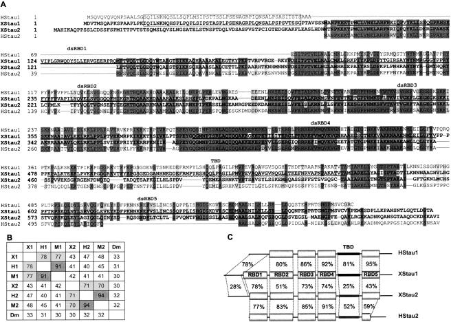
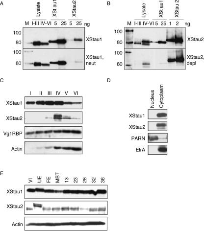
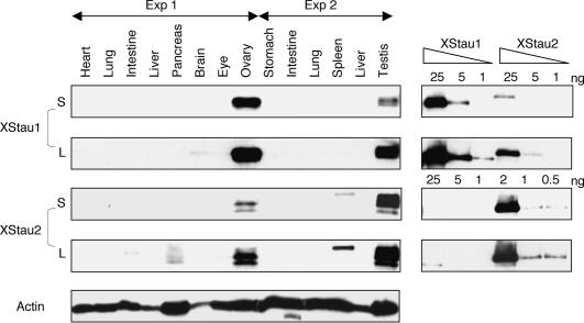
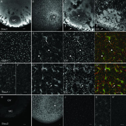
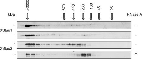
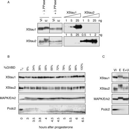
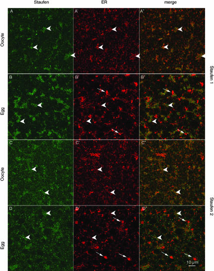
Localization of mRNA is an important way of generating early asymmetries in the developing embryo. In Drosophila, Staufen is intimately involved in the localization of maternally inherited mRNAs critical for cell fate determination in the embryo. We show that double-stranded RNA-binding Staufen proteins are present in the oocytes of a vertebrate, Xenopus, and are localized to the vegetal cytoplasm, a region where important mRNAs including VegT and Vg1 mRNA become localized. We identified two Staufen isoforms named XStau1 and XStau2, where XStau1 was found to be the principal Staufen protein in oocytes, eggs, and embryos, the levels of both proteins peaking during mid-oogenesis. In adults, Xenopus Staufens are principally expressed in ovary and testis. XStau1 was detectable throughout the oocyte cytoplasm by immunofluorescence and was concentrated in the vegetal cortical region from stage II onward. It showed partial codistribution with subcortical endoplasmic reticulum (ER), raising the possibility that Staufen may anchor mRNAs to specific ER-rich domains. We further showed that XStau proteins are transiently phosphorylated by the MAPK pathway during meiotic maturation, a period during which RNAs such as Vg1 RNA are released from their tight localization at the vegetal cortex. These findings provide evidence that Staufen proteins are involved in targeting and/or anchoring of maternal determinants to the vegetal cortex of the oocyte in Xenopus. The Xenopus oocyte should thus provide a valuable system to dissect the role of Staufen proteins in RNA localization and vertebrate development.


Figures

FIGURE 1.
FIGURE 1.
Xenopus Staufen 1 and 2 sequences. (A) XStau1 (AY705672) and XStau2 (BC046732) sequences, in bold, were aligned with human Stau163 (AL133174) and Stau262 (AK002152) using Clustal 1.82. Boundaries of gaps longer than 10 residues were refined by comparison to exon/intron junctions in human Stau genomic sequences. Dark gray shadowing indicates identity in at least three of the four sequences, and light gray indicates conserved changes. dsRBD1–5 domains in Xenopus Staufens, based on Drosophila dsRBD domains (St Johnston et al. 1992) are shown as bold lines, and the tubulin-binding domain (TBD) (Wickham et al. 1999) is shown as a wavy line. The N-terminal region conserved between XStau1 and human Stau163 is boxed. (B) Xenopus Staufens were aligned with the longest available versions of mammalian Stau proteins, namely, human Stau163 (H1, AL133174), mouse Stau155 (M1, BC012959), human Stau262 (H2, AK002152), and mouse Stau262 (M2, AF459099), as well as Drosophila Staufen (Dm, M69111) using Clustal 1.82, and scores of % homology between any two sequences are tabulated. (C) Schematic representation of Xenopus Stau1 and 2 and human Stau163 and Stau262. Identity scores for the domains indicated in A were obtained using the default settings of the GAP algorithm (GCG); the drawing is not to scale.
FIGURE 2.
FIGURE 2.
The levels of Xenopus Staufens peak in mid-oogenesis. (A,B) Validation of enrichment in XStau1- and XStau2-specific antibodies. Two cell equivalent mixed-stage oocytes lysates (I–III) and (IV–VI) and indicated amounts of recombinant Xenopus Staufen proteins were analyzed by Western blotting. (A) Top panel, using XStau1 antibody (1:10,000 dilution) and bottom panel XStau1 antibody (1:10,000) neutralized with XStau2 protein (XStau1, neutr). (B) Top panel, using XStau2 antibody (1:2500) and bottom panel XStau2 antibody (1:2500) depleted of XStau1 epitopes (XStau2, depl). Lane M, Magic Mark Western Standard molecular weight standards, in kDa. Donkey antirabbit secondary antibodies and the molecular weight standards were detected by ECL. (C) The levels of Xenopus Staufens peak in mid-oogenesis. Lysates were prepared from staged oocytes (I–VI) and two cell equivalents were analyzed by Western blotting with specific XStau1 and XStau2 antibodies. The oocyte samples were also probed with control Vg1RBP and actin antibodies. (D) XStau1 and 2 are cytoplasmic proteins. Nuclear and cytoplasmic fractions were prepared from stage VI oocytes and equivalent cell samples analyzed by Western blotting. The fractions were verified by PARN antibody, which detects the nuclear poly(A) nuclease and HuR antibody, which detects the cytoplasmic elrA protein. In C,D, XStau antibodies were used and verified against recombinant proteins as in panels A,B (data not shown). (E) XStau protein expression in oocytes, eggs, and embryos. Lysates from two cell equivalents of stage VI oocytes, unfertilized and fertilized eggs, as well as MBT and embryonic stages 13–36 were analyzed by Western blotting with specific XStau1 and XStau2 antibodies, verified by comparison with indicated amounts of recombinant proteins. A control blot of the same samples with actin antibodies confirmed consistent protein loading.
FIGURE 3.
FIGURE 3.
Xenopus Staufen expression in adults is largely confined to reproductive tissues. Samples were prepared from indicated adult tissues, in two independent experiments as detailed in Materials and Methods, and 40 μg of total protein was analyzed by Western blotting, alongside indicated amounts of recombinant proteins. Short (S, XStau1 10 sec, XStau2, 30 sec) and Long (L, XStau1 and XStau2, 10 min) exposures are shown. The blots were overprobed with anti-actin antibody.
FIGURE 4.
FIGURE 4.
Localization of XStau1 and XStau2 proteins in oocytes. Confocal images of oocytes stained with anti-XStau1 (AG) and anti-XStau2 (HK) antibodies. Stage I oocytes are shown in A,H; Stage II oocytes in B,C,I. All other images are of stage VI oocytes. Staining with both anti-Staufen antibodies was excluded from the mitochondrial cloud (MC) at stage I and from the site where it has joined the cortex at stage II–III (*). The images in D,FI are confocal sections through a vegetal subcortical layer of stage VI oocytes containing distinctive patches of ER, which lies about 5 μm beneath the oocyte surface. E is a more superficial confocal section of the same oocyte as G, taken at the level of the cortical ER network. C′, E′, and G′ show the ER distribution in oocytes costained with anti-GRP94. E″ and G″ are corresponding overlays of XStau1 (green) and GRP94 (red). Arrows in C,E,G point to sites of XStau1-ER colocalization; arrowheads in E indicate absence of localization at the level of the outer cortex. F′ and I′ are images of oocytes stained with antibodies pre-incubated with the corresponding protein, acquired with the same confocal settings as those of the oocytes stained with BSA-incubated antibodies in F and I. Scale bars 10 μm throughout.
FIGURE 5.
FIGURE 5.
XStau granules and particles in Xenopus oocyte lysates: 200 μL stage V/VI S10 extract was fractionated through a Superose 6 HR 10/30 column at a flow rate of 0.5 mL/min, and 0.3- mL fractions collected. Alternate fractions were separated by SDS-PAGE and visualized by Western blotting using antibodies as shown. Where indicated, lysate was treated with RNase A (167 μg/mL, 10 min, 20°C) prior to loading on the column. This treatment did not significantly affect the fractionation behavior of the Coomassie Blue-stainable proteins (data not shown). Elution volumes of size markers [Blue Dextran (> 2000 kDa), thyroglobulin (669 kDa), ferritin (440 kDa), catalase (232 kDa), aldolase (158 kDa), ovalbumin (43 kDa), and chymotryp-sinogen A (25 kDa)] are marked by arrows.
FIGURE 6.
FIGURE 6.
XStau1 and XStau2 are phosphorylated during meiotic maturation. (A) Lysates were prepared from stage VI oocytes and progesterone-matured eggs, and treated (+) or left untreated (−) with λ-phosphatase, and subsequently analyzed by Western blotting with specific XStau1 and XStau2 antibodies as in Figures 2 ▶ and 3 ▶. (B) Timing of phosphorylation during meiotic maturation. Random groups of 20 cells out of ~ 250 stage VI oocytes treated with progesterone were taken at the time of progesterone addition (T0) and when the indicated proportion of cells has undergone GVBD (%). Oocytes were then subjected to Western blot analysis with the indicated antibodies. (C) XStau1 and XStau2 are phosphorylated by the MAPK kinase pathway. Stage VI oocytes, some of which were preincubated with 50 μM U0126 for 4 h, were matured with progesterone. Samples of untreated oocytes (O), control matured eggs (E), and eggs matured in the presence of UO126 (E+U) were analyzed by western blotting with the indicated antibodies.
FIGURE 7.
FIGURE 7.
Staufen distribution in the vegetal region of oocytes and eggs. Confocal images of oocytes and progesterone-matured eggs stained with anti-XStau1 (A,C) and anti-XStau2 (B,D) antibodies. All samples were costained with anti-GRP 94 to visualize ER (images A′D′). A′D′ are the corresponding overlays of XStau (green) and GRP94 (red). These confocal sections were taken about 5 μm below the vegetal surface in a layer containing distinctive patches (arrowheads) containing XStau1, and to a lesser extent XStau2. In oocytes, these patches coincide closely with domains rich in ER. During maturation the ER reorganizes to form distinctive, brightly stained whorls of ER (white arrows in B,D), while ER is partially lost from the Staufen-containing patches. Scale bar, 10 μm.

Similar articles

Cited by

References

    1. Alarcon, V.B. and Elinson, R.P. 2001. RNA anchoring in the vegetal cortex of the Xenopus oocyte. J. Cell Sci. 114: 1731–1741. - PubMed
    1. Beckhelling, C., Chang, P., Chevalier, S., Ford, C., and Houliston, E. 2003. Pre-M Phase-promoting factor associates with annulate lamellae in Xenopus oocytes and egg extracts. Mol. Biol. Cell 14: 1125–1137. - PMC - PubMed
    1. Betley, J.N., Heinrich, B., Vernos, I., Sardet, C., Prodon, F., and Deshler, J.O. 2004. Kinesin II mediates Vg1 mRNA transport in Xenopus oocytes. Curr. Biol. 14: 219–224. - PubMed
    1. Braun, R.E. 1998. Post-transcriptional control of gene expression during spermatogenesis. Semin. Cell Dev. Biol. 9: 483–489. - PubMed
    1. Broadus, J., Fuerstenberg, S., and Doe, C.Q. 1998. Staufen-dependent localisation of prospero mRNA contributes to neuroblast daughter-cell fate. Nature 391: 792–795. - PubMed

Publication types

MeSH terms

LinkOut - more resources