

Review
doi: 10.1523/JNEUROSCI.3649-04.2004.
Mechanisms and roles of axon-Schwann cell interactions
Affiliations
- PMID: 15496660
- PMCID: PMC6730082
- DOI: 10.1523/JNEUROSCI.3649-04.2004
Item in Clipboard
Review
Mechanisms and roles of axon-Schwann cell interactions
J Neurosci.
.
No abstract available
Figures
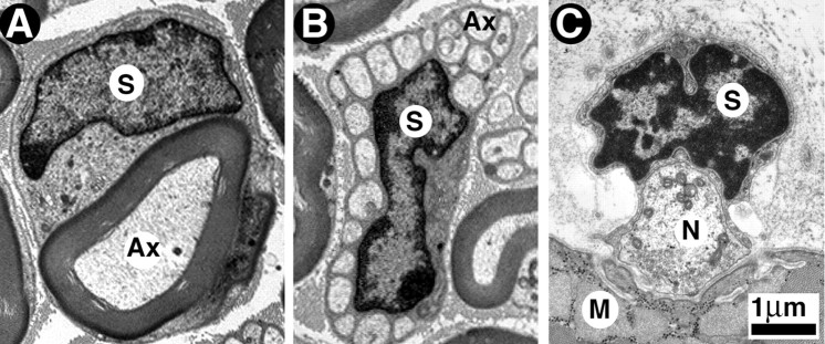
Myelinated, unmyelinated, and perisynaptic Schwann cells as seen with the electron microscope. A, Cross section of a myelinated axon of an adult mouse sciatic nerve. The myelin sheath (MS) surrounding the axon (Ax) and the Schwann cell nucleus (S) are clearly visible. B, Cross section of a bundle of unmyelinated axons of an adult mouse sciatic nerve. The Schwann cell forms the Remak bundle, a bouquet-like bundle of thin axons, each separated from its neighbor by thin cytoplasmic extensions of the Schwann cell. C, Cross section of a frog neuromuscular junction reveals three juxtaposed cellular elements: the perisynaptic Schwann cell, nerve terminal (N), and muscle fiber (M). The perisynaptic Schwann cell body (S indicates nucleus) and its processes cap the nerve terminal, but the processes do not wrap around the nerve terminal region facing acetylcholine receptors on muscle.
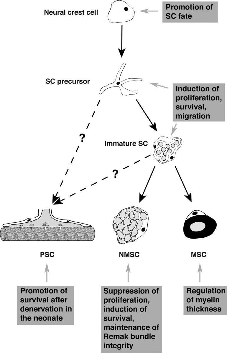
NRG1-erbB signaling and Schwann cell development. During development, neural crest cells give rise to Schwann cell precursors, which then develop into the three adult phenotypes: PSCs, NMSCs, or MSCs. Whereas, during their differentiation into MSCs and NMSCs, the precursors proceed through a stage called immature Schwann cell, the direct precursor of PSCs remains unknown. NRG1-erbB signaling regulates important aspects of Schwann cell biology at each step of their development (see boxed text).
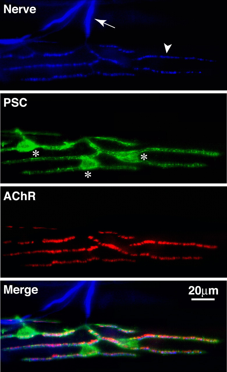
Perisynaptic Schwann cells at the neuromuscular junction. Frog skeletal muscle triple labeled with anti-neurofilament for axons (first panel, arrow) and synapsin I antibodies for nerve terminals (first panel, arrowhead), a monoclonal antibody, 2A12, for perisynaptic Schwann cells (second panel, the cell bodies are marked with *), and α-bungarotoxin for postsynaptic acetylcholine receptors (third panel). The merged image is shown in the fourth panel.
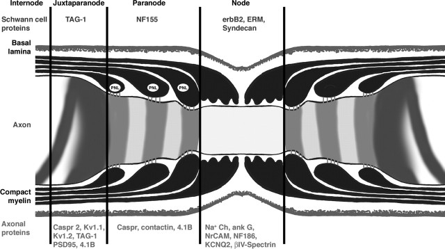
Molecular structure of the node of Ranvier. Longitudinal section through the nodal region of a peripheral myelinated axon showing the organization and composition of axonal and glial domains. The axon is covered by an MSC, which in turn is surrounded by a basal lamina. In the paranodal region, the myelin sheath forms a series of paranodal loops (PNL) that invaginate and appose the axon creating a septate-like structure. At the node, the outermost cytoplasmic extension of MSC contains numerous microvilli that contact the axolemma. Specific sets of proteins are enriched in each domain of both axon (red text) and Schwann cells (blue text).
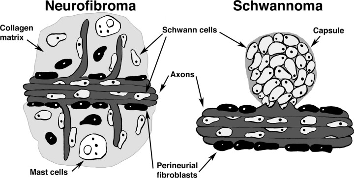
Schwann cell tumors. In neurofibromas, Schwann cells fibroblasts and perineurial cells are with in collagen-rich tumor matrix; axons and mast cells are common. Neurofibromas are not encapsulated, although the plexiform neurofibroma develops inside the perineurium (not shown). Schwannomas are encapsulated by a collagenous sheath and are made up almost entirely of S100β-positive Schwann cells, with little or no fibroblast involvement; axons are present only at the boundary of the tumor with the associated nerve trunk.
Similar articles
-
Development of axonal membrane specializations defines nodes of Ranvier and precedes Schwann cell myelin elaboration.Dev Biol. 1980 Oct;79(2):334-55. doi: 10.1016/0012-1606(80)90120-7. Dev Biol. 1980. PMID: 7429010 No abstract available.
-
Axonal domains: role for paranodal junction in node of Ranvier assembly.Curr Biol. 2008 Sep 23;18(18):R876-9. doi: 10.1016/j.cub.2008.07.070. Curr Biol. 2008. PMID: 18812088
-
[Axon and Schwann cells... so far away, so close].Rev Neurol (Paris). 2008 Dec;164(12):1057-62. doi: 10.1016/j.neurol.2008.10.003. Epub 2008 Nov 28. Rev Neurol (Paris). 2008. PMID: 19041107 Review. French.
-
Peripheral nerve regeneration.Neurosci Res. 1996 Jun;25(2):101-21. doi: 10.1016/0168-0102(96)01042-5. Neurosci Res. 1996. PMID: 8829147 Review.
-
The local differentiation of myelinated axons at nodes of Ranvier.Nat Rev Neurosci. 2003 Dec;4(12):968-80. doi: 10.1038/nrn1253. Nat Rev Neurosci. 2003. PMID: 14682359 Review.
Cited by
-
mTORC1 Activation by Loss of Tsc1 in Myelinating Glia Causes Downregulation of Quaking and Neurofascin 155 Leading to Paranodal Domain Disorganization.Front Cell Neurosci. 2018 Jul 12;12:201. doi: 10.3389/fncel.2018.00201. eCollection 2018. Front Cell Neurosci. 2018. PMID: 30050412 Free PMC article.
-
Myelin sheath structure and regeneration in peripheral nerve injury repair.Proc Natl Acad Sci U S A. 2019 Oct 29;116(44):22347-22352. doi: 10.1073/pnas.1910292116. Epub 2019 Oct 14. Proc Natl Acad Sci U S A. 2019. PMID: 31611410 Free PMC article.
-
Long-term modulation of the axonal refractory period.Eur J Neurosci. 2022 Oct;56(7):4983-4999. doi: 10.1111/ejn.15801. Epub 2022 Sep 1. Eur J Neurosci. 2022. PMID: 35999192 Free PMC article.
-
Neuronal merlin influences ERBB2 receptor expression on Schwann cells through neuregulin 1 type III signalling.Brain. 2014 Feb;137(Pt 2):420-32. doi: 10.1093/brain/awt327. Epub 2013 Dec 5. Brain. 2014. PMID: 24309211 Free PMC article.
-
Charcot-Marie-Tooth disease and intracellular traffic.Prog Neurobiol. 2012 Dec;99(3):191-225. doi: 10.1016/j.pneurobio.2012.03.003. Epub 2012 Mar 22. Prog Neurobiol. 2012. PMID: 22465036 Free PMC article. Review.
References
-
- Apostolski S, Sadiq SA, Hays A, Corbo M, Suturkova-Milosevic L, Chaliff P, Stefansson K, LeBaron RG, Ruoslahti E, Hays AP, Latov N (1994) Identification of Gal(beta 1-3)GalNAc bearing glycoproteins at the nodes of Ranvier in peripheral nerve. J Neurosci Res 38: 134-141. - PubMed
-
- Araque A, Parpura V, Sanzgiri RP, Haydon PG (1999) Tripartite synapses: glia, the unacknowledged partner. Trends Neurosci 22: 208-215. - PubMed
-
- Astrow SH, Qiang H, Ko CP (1998) Perisynaptic Schwann cells at neuromuscular junctions revealed by a novel monoclonal antibody. J Neurocytol 27: 667-681. - PubMed
-
- Auld DS, Colomar A, Belair EL, Castonguay A, Pinard A, Rousse I, Thomas S, Robitaille R (2003) Modulation of neurotransmission by reciprocal synapse-glial interactions at the neuromuscular junction. J Neurocytol 32: 1003-1015. - PubMed
Publication types
MeSH terms
Substances
Grants and funding
LinkOut - more resources
Full Text Sources