. 2004 Oct 20;24(42):9282-90.
doi: 10.1523/JNEUROSCI.2120-04.2004.

Astrocyte-associated fibronectin is critical for axonal regeneration in adult white matter

Affiliations

Astrocyte-associated fibronectin is critical for axonal regeneration in adult white matter

Veronica J Tom et al. J Neurosci. .

Abstract

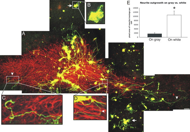
Although it has been suggested that astroglia guide pioneering axons during development, the cellular and molecular substrates that direct axon regeneration in adult white matter have not been elucidated. We show that although adult cortical neurons were only able to elaborate very short, highly branched, dendritic-like processes when seeded onto organotypic slice cultures of postnatal day 35 (P35) rat brain containing the corpus callosum, adult dorsal root ganglion (DRG) neurons were able to regenerate lengthy axons within the reactive glial environment of this degenerating white matter tract. The callosum in both P35 slices and adult rat brain was rich in fibronectin, but not laminin. Furthermore, the fibronectin was intimately associated with the intratract astrocytes. Blockade of fibronectin function in situ with an anti-fibronectin antibody dramatically decreased outgrowth of DRG neurites, suggesting that fibronectin plays an important role in axon regeneration in mature white matter. The critical interaction between regrowing axons and astroglial-associated fibronectin in white matter may be an additional factor to consider when trying to understand regeneration failure and devising strategies to promote regeneration.


Figures

Figure 1.
Figure 1.
Dissociated adult DRGs attach to organotypic slices containing white matter. A, Low-magnification bright-field image of an organotypic coronal slice containing the white matter tract the corpus callosum that had been in culture for 4 d. B, C, Dissociated GFP+ DRGs were seeded onto the callosal region of 1 DIV slices and fixed and stained at 4 DIV. The bright-field image of the slice is shown in B. The fluorescent image of the GFP+ neurons is shown in C. The white matter tract is denoted by the white lines. Note that many neurons were able to adhere to the white matter. Scale bars: A, 500 μm; B, C, 200 μm.
Figure 2.
Figure 2.
DRGs regenerate robustly on the corpus callosum. A, Confocal microscopy montage of GFP+ DRGs (green) on the GFAP+ astrocyte-rich corpus callosum (red). Insets are higher-magnification images of areas denoted by the white boxes. The DRGs were able to grow well on the callosum and extended long neurites (C, arrowhead) after 3 DIV; however, neurons on gray matter, which was virtually devoid of GFAP+ astrocytes, were able to extend only very short neurites (B, arrows in main figure A). Although much of the growth appeared to grossly follow the alignment of the host astrocyte network, there were numerous instances during which axons strayed and turned (C, D, arrowhead). E, Quantification of the disparity between outgrowth on gray and white matter. Neurites from DRGs seeded onto 1 DIV organotypic slice cultures and maintained for 3 DIV were traced digitally. Neurites originating from DRGs seeded on gray matter of the organotypic slice culture after 3 DIV were traced digitally and compared with those neurites that grew on the corpus callosum (n = 4 slices per group). The pixel areas of the tracings were quantified and analyzed statistically using a Student's t test. There were significantly shorter neurites per neuron on gray matter than on white matter (*p < 0.005). Scale bar, 100 μm.
Figure 3.
Figure 3.
Cortical cells do not regenerate well on white matter. Adult cortical neurons seeded onto the corpus callosum of P35 organotypic slice cultures were double stained for GFP (A, C, D, F, green) and the dendritic marker MAP-2 (B, C, E, F) after 8 DIV. Even after such a long time in culture, the neurons were able to extend only short neurites. Furthermore, all neurites were positive for MAP-2, suggesting that they were of a dendritic nature. Scale bar, 25 μm.
Figure 4.
Figure 4.
DRGs regenerate neurites despite the presence of myelin. A, B, An abundance of myelin was present in the corpus callosi of both 1 DIV slices (when neurons were seeded) and 4 DIV slices (when cultures were fixed and regeneration was assessed). C, D, Z-series of confocal microscope images revealed that although there were some GFP+ neurites that remained on the surface of the slice (D, green, arrowhead), the majority of regenerating neurites penetrated and grew ∼15-20 μm below the surface of the slice (D, yellow). There was plenty of myelin at this depth in the slice at both 1 and 4 DIV (C), suggesting that the regenerating neurites were growing among myelin as well as callosal astrocytes (D, red). Scale bar: A, B, 200 μm.
Figure 5.
Figure 5.
Regenerating neurites are only crudely aligned with host tract astrocytes. A, B, regenerating GFP+ neurites (green) generally followed host GFAP+ astrocytic processes in the callosum (red) and grew longitudinally (arrowheads). Alignment was not exact, however, and there were instances during which neurites wandered and jumped from one astrocytic process to another (B, asterisk). C, D, Regenerating neurites (green) in the cingulum grew radially away from the callosum, apparently guided by the astrocytes that were present there (arrows). Scale bar, 25 μm.
Figure 6.
Figure 6.
Fibronectin, not laminin, is expressed by astrocytes in the corpus callosum. A,B, Freshly fixed coronal slices stained for fibronectin (A) and laminin (B). Although laminin was visualized only along blood vessels, fibronectin was expressed all over the cortex, including the corpus callosum, demarcated by the blue dots. C, D, Perfused adult rat brain sections stained for fibronectin (C) and laminin (D). Again, the callosum was rich in fibronectin, whereas laminin was present only along blood vessels. E-G, A GFAP+ astrocyte in the callosum of a slice (F, G, red) was closely associated with fibronectin-expression patterns (E, G, arrows, green). Scale bars: A-D, 50 μm; E-G, 5 μm.
Figure 7.
Figure 7.
Polyclonal fibronectin (FN) and laminin (LN) antibodies are function blocking. A-C, G, Dissociated, adult DRGs were plated onto fibronectin-coated (A, B) or laminin-coated coverslips (C) and treated with control rabbit IgG (A), rabbit anti-fibronectin (B), or rabbit anti-laminin (C) for 3 d and then stained for β-tubulin III. Although RIgG had no effect on neurite outgrowth, anti-fibronectin virtually abolished all outgrowth on fibronectin, and anti-laminin greatly diminished outgrowth on laminin. The longest neurite of randomly chosen neurons on either FN- or LN-coated coverslips treated with either RIgG or the appropriate antibody (n = 12 per group) was measured and compared (G). There was a statistical difference between the RIgG and anti-fibronectin treatments on neurites growing on fibronectin (*p < 0.00001), and between RIgG and anti-laminin treatments on neurites growing on laminin (**p < 0.00000001). D-F, To control for antibody-mediated toxicity, adult DRGs were plated onto poly-lysine-coated coverslips and treated with RIgG (D), anti-fibronectin (E), or anti-laminin (F) for 3 d. Neurons were able to extend long neurites with any of the three treatments, suggesting that the antibodies were not toxic, and both antibodies were able to specifically block outgrowth mediated by their respective molecules. Scale bar, 50 μm.
Figure 8.
Figure 8.
Anti-fibronectin, but not anti-laminin, decreases regeneration on white matter. After neuron seeding, the slice cultures were treated with control RIgG (A), anti-fibronectin (B), or anti-laminin (C) for 3 d. GFP immunohistochemistry is shown in A, B, and D. Blood vessels were also stained because the anti-rabbit secondary used to stain the GFP+ neurites also recognized the anti-laminin and anti-fibronectin antibodies. Although there was extensive outgrowth along the corpus callosum in RIgG- and anti-laminin-treated slices, there was a diminished amount of outgrowth in anti-fibronectin-treated slices. C, High-magnification image of a fibronectin-treated slice double stained for GFP (green) and GFAP (red). Although the GFAP+ astrocytes in the fibronectin-treated slices appeared smaller than in RIgG- or anti-laminin-treated slices (compare with Figs. 2 and 5 A, B), GFP+ neurites (green) were not able to grow well even on an intact, longitudinal astrocytic terrain (arrowhead). E, Outgrowth on white matter of slices treated with RIgG (n = 10), anti-fibronectin (n = 10), or anti-laminin (n = 5) was digitally traced. The pixel areas of the tracings were quantified and statistically compared using a Student's t test. Although there was no significant difference between RIgG- and anti-laminin-treated slices, anti-fibronectin statistically decreased outgrowth when compared with the RIgG-treated controls (*p < 0.0001). These findings suggest that astrocyte-associated fibronectin plays a critical role in regeneration on the corpus callosum. Scale bars: A, B, D, 100 μm; C, 25 μm.

Similar articles

Cited by

References

    1. Adachi K, Yamauchi K, Bernaudin JF, Fouret P, Ferrans VJ, Crystal RG (1988) Evaluation of fibronectin gene expression by in situ hybridization: differential expression of the fibronectin gene among populations of human alveolar macrophages. Am J Pathol 133: 193-203. - PMC - PubMed
    1. Ard MD, Bunge MB, Wood PM, Schachner M, Bunge RP (1991) Retinal neurite growth on astrocytes is not modified by extracellular matrix, anti-L1 antibody, or oligodendrocytes. Glia 4: 70-82. - PubMed
    1. Asher RA, Morgenstern DA, Fidler PS, Adcock KH, Oohira A, Braistead JE, Levine JM, Margolis RU, Rogers JH, Fawcett JW (2000) Neurocan is upregulated in injured brain and in cytokine-treated astrocytes. J Neurosci 20: 2427-2438. - PMC - PubMed
    1. Bandtlow C, Zachleder T, Schwab ME (1990) Oligodendrocytes arrest neurite growth by contact inhibition. J Neurosci 10: 3837-3848. - PMC - PubMed
    1. Baron-Van Evercooren A, Kleinman HK, Ohno S, Marangos P, Schwartz JP, Dubois-Dalcq ME (1982) Nerve growth factor, laminin, and fibronectin promote neurite growth in human fetal sensory ganglia cultures. J Neurosci Res 8: 179-193. - PubMed

Publication types

MeSH terms