Localizing P300 generators in visual target and distractor processing: a combined event-related potential and functional magnetic resonance imaging study
- PMID: 15496671
- PMCID: PMC6730097
- DOI: 10.1523/JNEUROSCI.1897-04.2004
Localizing P300 generators in visual target and distractor processing: a combined event-related potential and functional magnetic resonance imaging study
Abstract
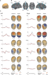
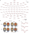
Constraints from functional magnetic resonance imaging (fMRI) were used to identify the sources of the visual P300 event-related potential (ERP). Healthy subjects performed a visual three-stimulus oddball paradigm with a difficult discrimination task while fMRI and high-density ERP data were acquired in separate sessions. This paradigm allowed us to differentiate the P3b component of the P300, which has been implicated in the detection of rare events in general (target and distractor), from the P3a component, which is mainly evoked by distractor events. The fMRI-constrained source model explained >99% of the variance of the scalp ERP for both components. The P3b was mainly produced by parietal and inferior temporal areas, whereas frontal areas and the insula contributed mainly to the P3a. This source model reveals that both higher visual and supramodal association areas contribute to the visual P3b and that the P3a has a strong frontal contribution, which is compatible with its more anterior distribution on the scalp. The results point to the involvement of distinct attentional subsystems in target and distractor processing.
Figures


Similar articles
-
Auditory P3a and P3b neural generators in schizophrenia: An adaptive sLORETA P300 localization approach.Schizophr Res. 2015 Dec;169(1-3):318-325. doi: 10.1016/j.schres.2015.09.028. Epub 2015 Oct 9. Schizophr Res. 2015. PMID: 26481687
-
[Brain electrical source analysis of the response to visual target and distractor stimuli].Sheng Wu Yi Xue Gong Cheng Xue Za Zhi. 2012 Oct;29(5):933-40. Sheng Wu Yi Xue Gong Cheng Xue Za Zhi. 2012. PMID: 23198438 Chinese.
-
Attentional systems in target and distractor processing: a combined ERP and fMRI study.Neuroimage. 2004 Jun;22(2):530-40. doi: 10.1016/j.neuroimage.2003.12.034. Neuroimage. 2004. PMID: 15193581
-
The neurophysiology of P 300--an integrated review.Eur Rev Med Pharmacol Sci. 2015 Apr;19(8):1480-8. Eur Rev Med Pharmacol Sci. 2015. PMID: 25967724 Review.
-
Updating P300: an integrative theory of P3a and P3b.Clin Neurophysiol. 2007 Oct;118(10):2128-48. doi: 10.1016/j.clinph.2007.04.019. Epub 2007 Jun 18. Clin Neurophysiol. 2007. PMID: 17573239 Free PMC article. Review.
Cited by
-
Causality in the association between P300 and alpha event-related desynchronization.PLoS One. 2012;7(4):e34163. doi: 10.1371/journal.pone.0034163. Epub 2012 Apr 12. PLoS One. 2012. PMID: 22511933 Free PMC article.
-
Changes in P3b Latency and Amplitude Reflect Expertise Acquisition in a Football Visuomotor Learning Task.PLoS One. 2016 Apr 25;11(4):e0154021. doi: 10.1371/journal.pone.0154021. eCollection 2016. PLoS One. 2016. PMID: 27111898 Free PMC article.
-
Acute and subchronic effects of amitriptyline on processing capacity in neuropathic pain patients using visual event-related potentials: preliminary findings.Psychopharmacology (Berl). 2006 Jan;183(4):462-70. doi: 10.1007/s00213-005-0204-3. Epub 2005 Nov 15. Psychopharmacology (Berl). 2006. PMID: 16292592 Clinical Trial.
-
Timing and sequence of brain activity in top-down control of visual-spatial attention.PLoS Biol. 2007 Jan;5(1):e12. doi: 10.1371/journal.pbio.0050012. PLoS Biol. 2007. PMID: 17199410 Free PMC article.
-
Mental training affects distribution of limited brain resources.PLoS Biol. 2007 Jun;5(6):e138. doi: 10.1371/journal.pbio.0050138. PLoS Biol. 2007. PMID: 17488185 Free PMC article.
References
-
- Anderer P, Pascual-Marqui RD, Semlitsch HV, Saletu B (1998) Differential effects of normal aging on sources of standard N1, target N1 and target P300 auditory event-related brain potentials revealed by low resolution electromagnetic tomography (LORETA). Electroencephalogr Clin Neurophysiol 108: 160-174. - PubMed
-
- Ardekani BA, Choi SJ, Hossein-Zadeh GA, Porjesz B, Tanabe JL, Lim KO, Bilder R, Helpern JA, Begleiter H (2002) Functional magnetic resonance imaging of brain activity in the visual oddball task. Brain Res Cogn Brain Res 14: 347-356. - PubMed
-
- Baudena P, Halgren E, Heit G, Clarke JM (1995) Intracerebral potentials to rare target and distractor auditory and visual stimuli. III. Frontal cortex. Electroencephalogr Clin Neurophysiol 94: 251-264. - PubMed
-
- Bledowski C, Prvulovic D, Goebel R, Zanella FE, Linden DE (2004) Attentional systems in target and distractor processing: a combined ERP and fMRI study. NeuroImage 22: 530-540. - PubMed
-
- Corbetta M, Shulman GL (2002) Control of goal-directed and stimulus-driven attention in the brain. Nat Rev Neurosci 3: 201-215. - PubMed
Publication types
MeSH terms
LinkOut - more resources
Full Text Sources
Miscellaneous
