. 2004 Oct 20;24(42):9391-404.
doi: 10.1523/JNEUROSCI.3314-04.2004.

A functional role of postsynaptic density-95-guanylate kinase-associated protein complex in regulating Shank assembly and stability to synapses

Affiliations

A functional role of postsynaptic density-95-guanylate kinase-associated protein complex in regulating Shank assembly and stability to synapses

Stefano Romorini et al. J Neurosci. .

Abstract

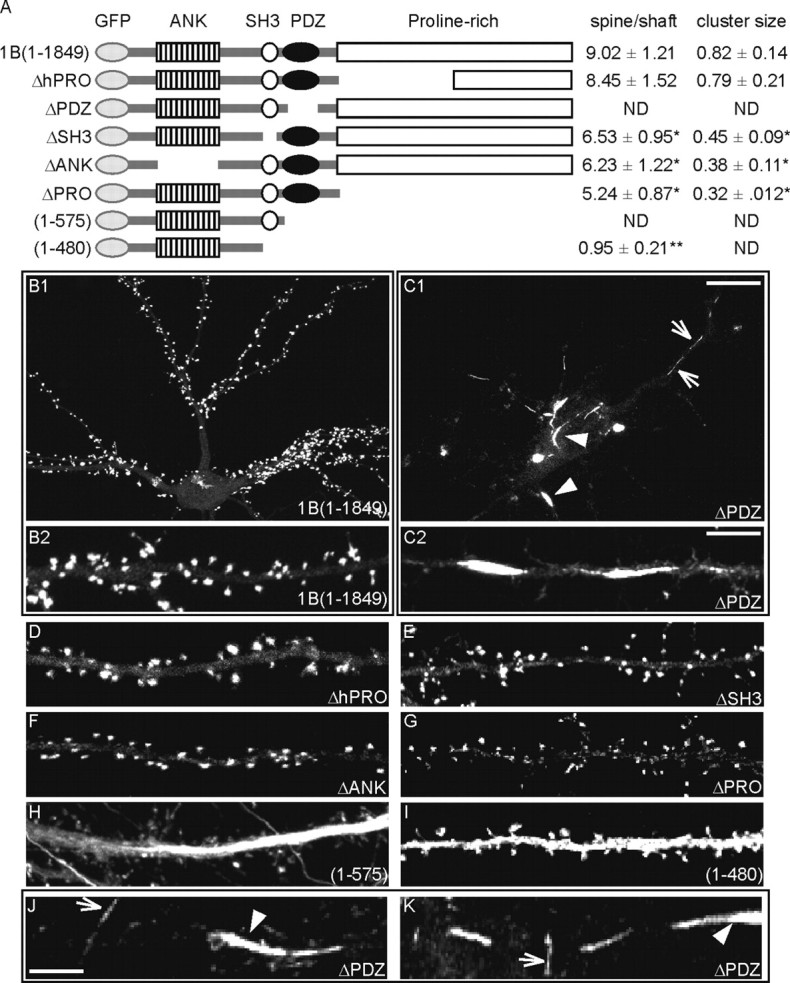
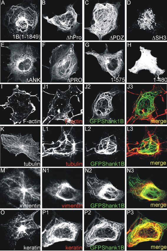
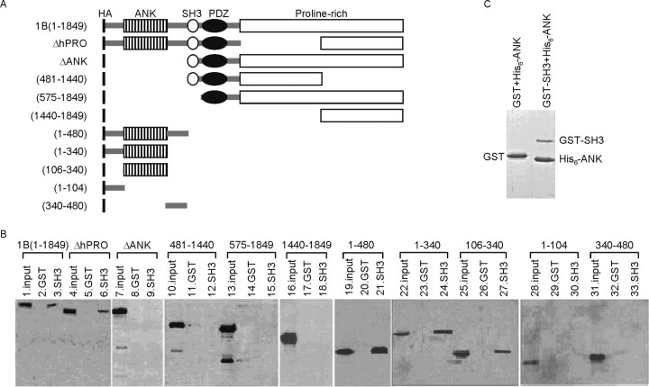
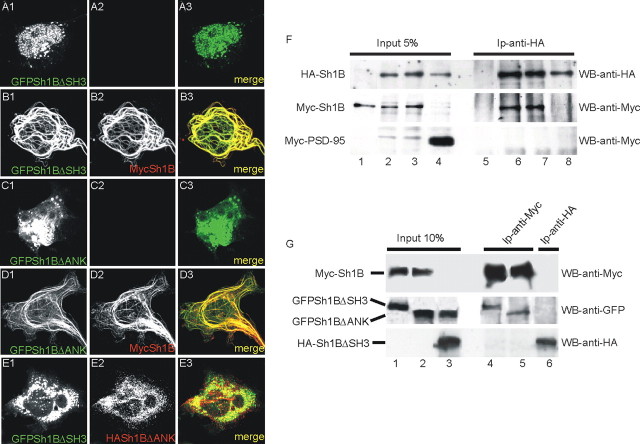
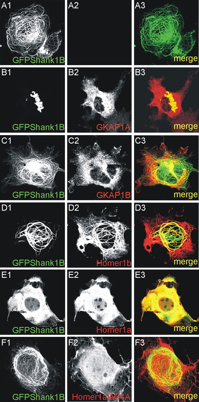
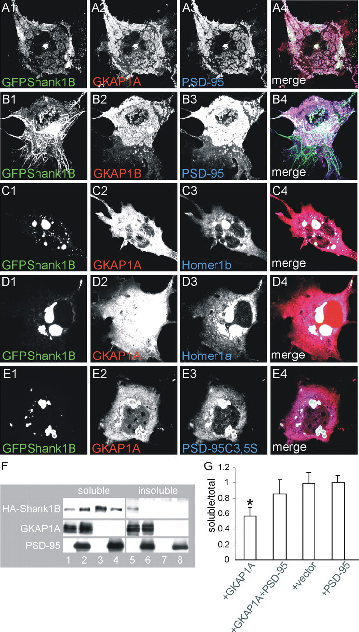
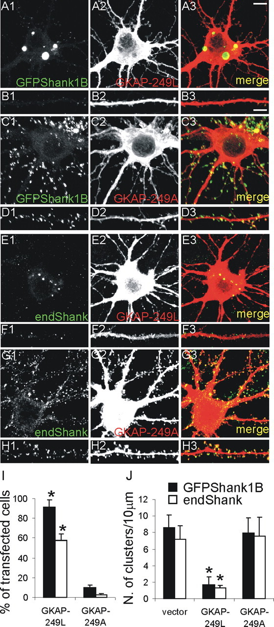
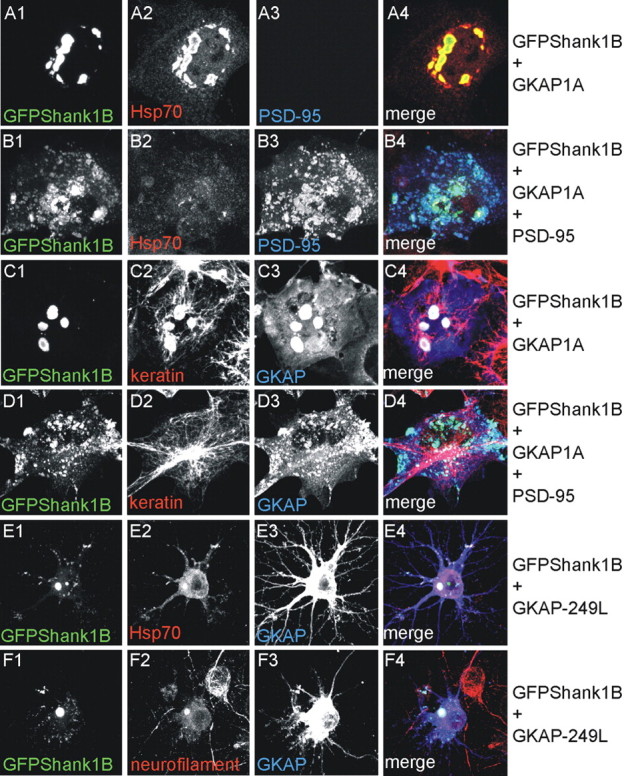
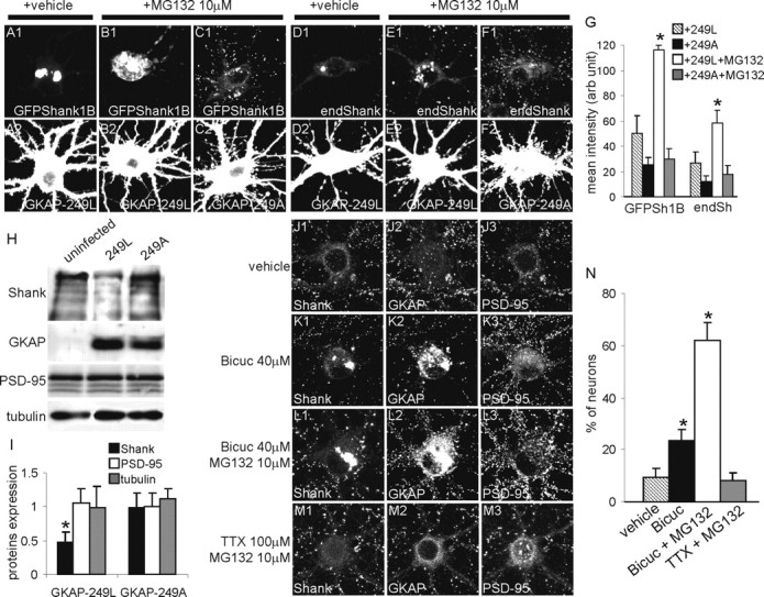
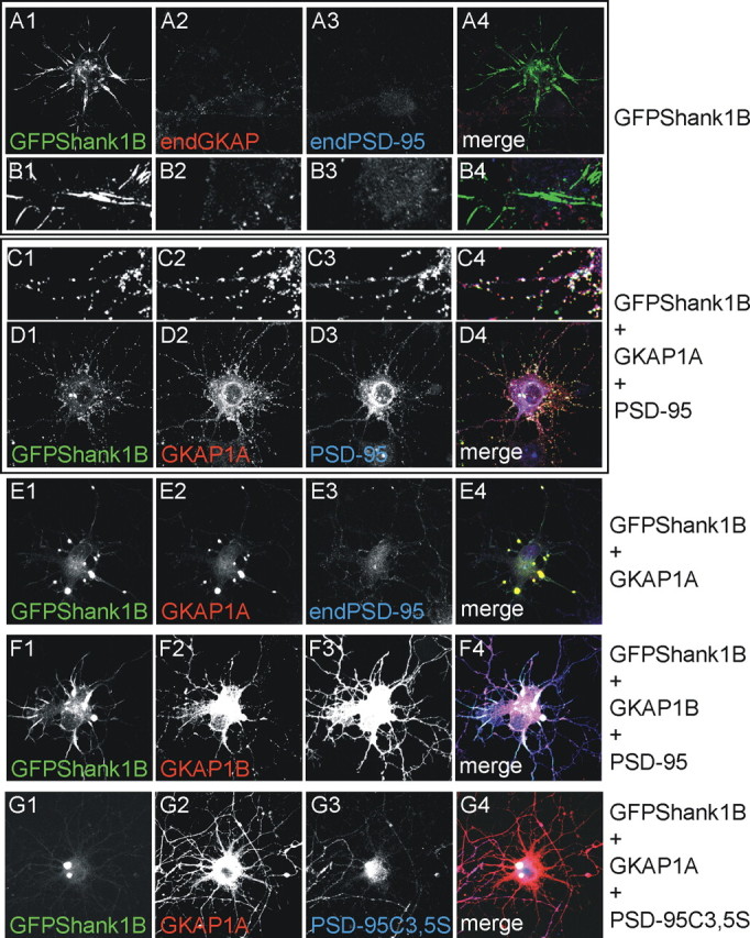
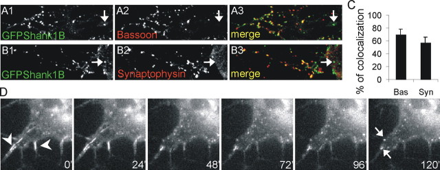
Postsynaptic density (PSD) proteins include scaffold, cytoskeletal, and signaling proteins that structurally and functionally interact with glutamate receptors and other postsynaptic membrane proteins. The molecular mechanisms regulating the assembly of PSD proteins and their associations with synapses are still widely unknown. We investigated the molecular mechanisms of Shank1 targeting and synapse assembly by looking at the function of guanylate kinase-associated protein (GKAP) and PSD-95 interactions. Shank1 when it is not associated to GKAP, which binds to the Shank PSD-95-Discs Large-zona occludens-1 domain, forms filamentous and fusiform structures in which the Src homology 3 domain specifically interacts with the ankyrin repeat domain, thus allowing its multimerization via a novel form of intermolecular interaction. Surprisingly, in both COS-7 cells and hippocampal neurons, GKAP forms insoluble aggregates with Shank that colocalize with heat shock protein 70 and neurofilaments, two markers of the aggresomes in which misfolded proteins accumulate. However, the two proteins are organized in clusters in COS cells and synaptic clusters in neurons when both are overexpressed and associated with wild-type PSD-95, but not with palmitoylation-deficient PSD-95. Synaptic activity in neurons induces the formation of Shank and GKAP intracellular aggregation and degradation. Similarly, the overexpression of a GKAP mutant that is incapable of binding PSD-95 induces Shank aggregation and degradation in neurons. Our data suggest a possible functional and structural role of the PSD-95-GKAP complex in Shank and PSD protein assembly and stability to synapses.


Figures

Figure 1.
Figure 1.
The PDZ domain is necessary for Shank1B targeting to spine. A, Diagram of the wild-type and mutant constructs of Shank1B used in this study (all GFP-tagged at the N terminus). The various domains are indicated. ANK, Ankyrin repeats. For each construct, the spine-shaft signal intensity and cluster size ratios (value ± SEM) were calculated as indicated on the right. ND, Not detected. *p < 0.05 versus GFPShank1B; **p < 0.01 versus GFPShank1B. B-K, Neurons were transfected with GFP-tagged wild-type Shank1B or mutant Shank constructs (as indicated in each panel) on DIV10-11 and fixed on DIV17-18; the images were acquired by means of confocal microscope in the GFP channel. B2 shows enlargements of a neuron dendrite transfected with GFPShank1B; C2, J, and K show enlargements of dendrites from neurons transfected with GFPShank1BΔPDZ. The constructs deleted from the PDZ domain, but with the SH3 and ANK domains, form fusiform (arrowheads) or filiform (arrows) aggregates in dendrites (C1, C2, H, J, K). Scale bars: (in C1) B1, C1, 10 μm; (in C2) B2, C2, D-I, 5 μm; (in J) J, K, 2.5 μm.
Figure 2.
Figure 2.
GFP-Shank forms filaments in COS cells. A-H, COS cells were transfected with the same constructs as those shown in Figure 1 and fixed 2 d after transfection. All of the constructs formed filaments in COS cells except the mutants deleted of the ankyrin or SH3 domains (D, E, H). I-P, COS cells were transfected or not with GFP-Shank1B and stained for F-actin (I, J1-J3), tubulin (K,L1-L3), vimentin (M,N1-N3), or keratin (O,P1-P3). The distribution of tubulin, vimentin, and keratin was modified by GFP-Shank1B: see K versus L1 for tubulin, M versus N1 for vimentin, and O versus P1-P3 for keratin.
Figure 3.
Figure 3.
The Shank1 SH3 domain binds to the ankyrin repeat domain (ANK). A, Diagram of the wild-type and mutant constructs of Shank1B (all HA-tagged at the N terminus) used in this study. B, Extracts of HEK cells transfected with wild-type Shank1B or mutant Shank constructs (as indicated above the Western blot) were incubated with GST or GST-SH3, and the pull-down proteins were revealed using HA antibody. In the input lane, ∼10% of the total extract used for the pull-down was loaded. C, GST-SH3, but not GST alone, can pull down purified His6-ANK domains.
Figure 4.
Figure 4.
GFPSh1BΔSH3 and GFPSh1BΔANK form filaments in COS cells only when coexpressed with full-length Shank1B. A, B, COS cells were transfected with GFPSh1BΔSH3 or GFPSh1BΔANK plus or minus Myc-Shank1B and stained for Myc in red; GFPSh1BΔSH3 and GFPSh1BΔANK was visualized on the GFP channel. E, COS cells were transfected with GFPSh1BΔDANK plus HA-Sh1BΔSH3 and stained for HA in red; GFPSh1BΔANK and HA-Sh1BΔSH3 did not colocalize when coexpressed in COS cells. F, COS cells were transfected with Myc-Shank1B alone (lanes 1,5), Myc-Shank1B plus HA-Shank1B (lanes 2, 3, 6, 7), and HA-Shank1B plus Myc-PSD-95 (lanes 4, 8). Extracts immunoprecipitated with HA antibodies were stained with HA and Myc antibodies, as indicated on the right. G, COS cells were transfected with Myc-Shank1B plus GFPShank1ΔSH3 (lanes 1,4), Myc-Shank1B plus GFPShank1ΔANK (lanes 2,5) or HA-Shank1ΔSH3 plus GFPShank1ΔANK (lanes 3,6). Extracts immunoprecipitated with Myc (lanes 4, 5) or HA (lane 6) antibodies were stained with Myc, GFP, or HA antibodies, as indicated on the right.
Figure 5.
Figure 5.
GKAP and Homer1a interfere with Shank1B filament distribution in COS cells. A-F, COS cells were transfected with GFP-Shank1B, alone or in combination with GKAP1A (a splice variant of GKAP that binds to Shank), GKAP1B (a splice variant of GKAP that does not bind to Shank), Homer1b, Homer1a, or Homer1aW24A, as indicated in each panel.
Figure 6.
Figure 6.
Only GFP-Shank1B, GKAP1A, and wild-type PSD-95 are distributed in clusters when coexpressed in COS cells. A, B, COS cells were triple-transfected with GFP-Shank1B plus GKAP1A or GKAP1B and PSD-95, as indicated in each panel. C-E, COS cells were triple-transfected with GFP-Shank1B plus GKAP1A or GKAP1B and Homer1b or Homer1a or PSD-95C3,5S, as indicated in each panel. The individual channels are shown in grayscale for better resolution; merge is shown in the panels on the right. F, G, HA-Shank1B solubility to detergent was tested in COS cells in the presence of GKAP1A alone or GKAP1A plus PSD-95. HA-Shank1B was soluble when transfected alone (F, lanes 3, 7, G) but became at least 50% insoluble when cotransfected with GKAP1A (F, lanes 1, 5, G). Shank1B solubility was recovered when GKAP1A was cotransfected with PSD-95 (F, lanes 2, 6, G). The graph bars indicate the mean values (±SEM) obtained from at least two independent experiments. *p < 0.05 versus all the other transfection combinations.
Figure 7.
Figure 7.
The GKAP1A-C-terminal (deleted of the N-terminal PSD-95 binding domain, GKAP-249L) induces aggregation in the cell body of transfected GFP-Shank1B and endogenous Shank. A-D, Neurons were transfected on DIV11 with Shank1B plus GKAP-249L or GKAP-249A (with a point mutation that abolishes binding to the Shank PDZ domain) as indicated in each panel, and stained on DIV16; B1-B3 and D1-D3, respectively, show higher magnifications of neuron dendrites from A1-A3 and C1-C3. E-H, Neurons were transfected on DIV11 with GKAP-249L or GKAP-249A, as indicated in the panel, and stained for GKAP and endogenous Shank on DIV16; F1-F3 and H1-H3, respectively, show higher magnifications of neuron dendrites from E1-E3 and G1-G3. I, Quantification of the numbers of neurons with intracellular aggregates formed by GFP-Shank1B and endogenous Shank after transfection with the indicated constructs (mean values ± SEM). J, Density of clusters of GFP-Shank1B and endogenous Shank per 10 μm of dendrite length (mean values ± SEM) in neurons transfected with the indicated constructs. At least 15 transfected neurons were quantified for each GKAP mutant construct. Scale bars: (in A3) A1-A3, C1-C3, E1-E3, G1-G3, 10 μm; (in B3) B1-B3, D1-D3, F1-F3, H1-H3, 5 μm. *p < 0.01 versus vector or GKAP-249A.
Figure 8.
Figure 8.
GKAP-Shank aggregates can be considered aggresomes. A-D, As indicated on the right, COS cells were transfected with GFPShank1B and GKAP1A (A, C) or GFPShank1B, GKAP1A, and PSD-95 (B, D) and stained for GFP (A1-D1), Hsp70 (A2, B2), PSD-95 (A3, B3), keratin (C2, D2), and GKAP (C3, D3). As shown in A1-A4, intracellular GFPShank1B-GKAP aggregates can be specifically stained with Hsp70 antibodies. The merge is shown in the panels on the right (A-D4). E, F, As indicated on the right, neurons were transfected on DIV10 with GFPShank1B plus GKAP-249L and stained on DIV15 for GFP (E1, F1), Hsp70 (E2), neurofilament (F2), and GKAP (E3, F3). The merge is shown in the panels on the right (E4, F4). Also in neurons, intracellular GFPShank1B-GKAP aggregates can be specifically stained with Hsp70 or neurofilament antibodies.
Figure 9.
Figure 9.
GKAP-249L induces partial degradation of GFPShank1B and endogenous Shank in neurons by means of proteasome degradation. A-F, DIV10 neurons were transfected with GFPShank1B plus GKAP-249L (A, B), GFPShank1B plus GKAP-249A (C), GKAP-249L alone (D, E), or GKAP-249A alone (F) and stained on DIV15 for GFP (A-C1), Shank (D-F1), and GKAP (A-F2). To test whether proteasomes are involved in this process, we treated the neurons with 10 μm of the MG132 proteasome inhibitor for 12 hr. G, The graph indicates the mean fluorescence intensity (arbitrary units ± SE) of the cell bodies of MG132-treated or untreated neurons. At least 10 neurons were measured at each experimental point; *p < 0.01 versus GKAP-249L without MG132. H, Neurons were infected at DIV5 with a lentivirus vector expressing GKAP-249L or GKAP-249A and lysated on DIV14. The expression of endogenous Shank, PSD-95, and tubulin was measured by Western blotting and quantified as shown in I. I, The graph indicates the mean value ± SE of the band intensity ratios between infected and uninfected neurons. The values were obtained from three independent experiments; *p < 0.05 versus GKAP249A. J-M, Bicuculline stimulation induces the formation of endogenous Shank and GKAP aggregates in cell bodies. The neurons were treated for 24 hr as indicated on the left of each panel and then stained for endogenous Shank (J1-M1), GKAP (J2-M2), and PSD-95 (J3-M3). Intracellular aggregates formed by endogenous Shank and GKAP (but not PSD-95) are visible in the neurons treated with bicuculline or bicuculline plus MG132. N, The graph indicates the mean ± SE percentage of neurons with intracellular aggregates. The data were obtained by counting at least 100 neurons from six coverslips obtained from three independent experiments for each treatment; *p < 0.01 versus vehicle and TTX plus MG132.
Figure 10.
Figure 10.
In young neurons, GFPShank1B is localized in synaptic-like clusters only if cotransfected with GKAP and PSD-95. A-D, As indicated on the right, the neurons were transfected on DIV4-5 with GFPShank1B alone (A1-A4, B1-B4), GFPShank1B and GKAP1A (E1-E4), GFPShank1B, GKAP1A, and PSD-95 (C1-C4, D1-D4), GFPShank1B, GKAP1B, and PSD-95 (F1-F4) or GFPShank1B, GKAP1B, and PSD-95C3,5S (G1-G4), and fixed on DIV6-7. The transfected neurons were labeled for GFP (A-G1), GKAP (A-G2), and PSD-95 (A-G3); the merge is shown in the panels on the right (A-G4). B1-B4 and C1-C4, respectively, show enlargements of A1-A4 and D1-D4.
Figure 11.
Figure 11.
In young neurons, the majority of the clusters formed by GFPShank1B, GKAP, and PSD-95 colocalize with presynaptic markers. A, B, The neurons were transfected on DIV4-5 with GFPShank1B, GKAP1A, and PSD-95 and fixed on DIV6-7. The transfected neurons were labeled for GFP (A1, B1), bassoon (A2), or synaptophysin (B2); the merge is shown in the panels on the right (A3, B3). C, The graph shows the mean ± SE percentage of GFPShank1B clusters that colocalize with bassoon or synaptophysin. The data were obtained by counting at least 20 neurons from six coverslips obtained from three independent experiments for each antibody. D, DIV5 neurons were transfected with GFPShank1B and imaged on DIV8-10 using a CCD camera on an inverted microscope for 2-4 hr at 37°C in a 5% CO2 atmosphere. The images show the pictures acquired after 0, 24, 48, 72, 96, and 120 min. The first frame of the figure shows two small filaments formed by GFPShank1B (arrowheads). These filaments progressively became reorganized in small aggregates, as also indicated by the two arrows in the last frame acquired 120 min after the first.

Similar articles

Cited by

References

    1. Beneken J, Tu JC, Xiao B, Nuriya M, Yuan JP, Worley PF, Leahy DJ (2000) Structure of the Homer EVH1 domain-peptide complex reveals a new twist in polyproline recognition. Neuron 26: 143-154. - PubMed
    1. Bockmann J, Kreutz MR, Gundelfinger ED, Bockers TM (2002) ProSAP/Shank postsynaptic density proteins interact with insulin receptor tyrosine kinase substrate IRSp53. J Neurochem 83: 1013-1017. - PubMed
    1. Boeckers TM, winter C, Smalla KH, Kreutz MR, Bockmann J, Seidenbecher C, Garner CC, Gundelfinger ED (1999) Proline-rich synapse-associated proteins ProSAP1 and ProSAP2 interact with synaptic proteins of the SAPAP/GKAP family. Biochem Biophys Res Commun 264: 247-252. - PubMed
    1. Boeckers TM, Bockmann J, Kreutz MR, Gundelfinger ED (2002) ProSAP/Shank proteins - a family of higher order organizing molecules of the postsynaptic density with an emerging role in human neurological disease. J Neurochem 81: 903-910. - PubMed
    1. Bonaglia MC, Giorda R, Borgatti R, Felisari G, Gagliardi C, Selicorni A, Zuffardi O (2001) Disruption of the ProSAP2 gene in a t(12;22)(q24.1; q13.3) is associated with the 22q13.3 deletion syndrome. Am J Hum Genet 69: 261-268. - PMC - PubMed

Publication types

MeSH terms

Substances

LinkOut - more resources