. 2004 Oct 20;24(42):9425-33.
doi: 10.1523/JNEUROSCI.2457-04.2004.

Rapid, activity-induced increase in tissue plasminogen activator is mediated by metabotropic glutamate receptor-dependent mRNA translation

Affiliations

Rapid, activity-induced increase in tissue plasminogen activator is mediated by metabotropic glutamate receptor-dependent mRNA translation

Chan Y Shin et al. J Neurosci. .

Abstract

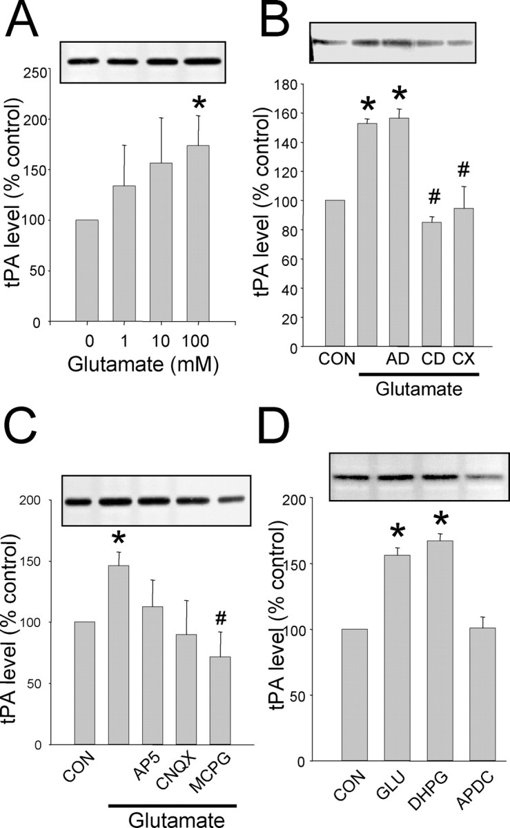
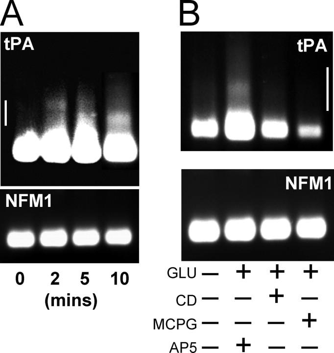
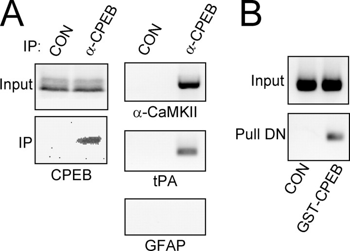
Long-term synaptic plasticity is both protein synthesis-dependent and synapse-specific. Therefore, the identity of the newly synthesized proteins, their localization, and mechanism of regulation are critical to our understanding of this process. Tissue plasminogen activator (tPA) is a secreted protease required for some forms of long-term synaptic plasticity. Here, we show tPA activity is rapidly increased in hippocampal neurons after glutamate stimulation. This increase in tPA activity corresponds to an increase in tPA protein synthesis that results from the translational activation of mRNA present at the time of stimulation. Furthermore, the mRNA encoding tPA is present in dendrites and is rapidly polyadenylated after glutamate stimulation. Both the polyadenylation of tPA mRNA and the subsequent increase in tPA protein is dependent on metabotropic glutamate receptor (mGluR) activation. A similar mGluR-dependent increase in tPA activity was detected after stimulation of a synaptic fraction isolated from the hippocampus, suggesting tPA synthesis is occurring in the synaptodendritic region. Finally, we demonstrate that tPA mRNA is bound by the mRNA-binding protein CPEB (cytoplasmic polyadenylation element binding protein-1), a protein known to regulate mRNA translation via polyadenylation. These results indicate that neurons are capable of synthesizing a secreted protein in the synaptic region, that mGluR activation induces mRNA polyadenylation and translation of specific mRNA, and suggest a model for synaptic plasticity whereby translational regulation of an immediate early gene precedes the increase in gene transcription.


Figures

Figure 1.
Figure 1.
Expression of tPA protein localizes to dendrites and synapses. A, Western blot of rat hippocampal homogenate probed with an affinity-purified anti-tPA antibody recognizes a single band of ∼68 kDa (arrow). Molecular weight markers shown on the left. B, tPA immunoreactivity (red) is detected in the cell body and distributed in puncta on hippocampal neurons in culture. This distribution is similar to that of synaptophysin (green). C, Images in B overlaid with each other and on the bright-field phase image showing dendritic distribution of tPA. Yellow puncta indicate a colocalization of tPA with synaptophysin (arrows). Box indicates an area of the cell shown at higher magnification in the right panel showing tPA protein also distributed in small puncta throughout the dendrite (arrowheads). Scale bars: B, C, left panel, 20 μm; C, right panel, 5 μm.
Figure 2.
Figure 2.
Stimulation of tPA synthesis by glutamate in hippocampal neurons in culture. A, Hippocampal neurons in culture (14-16 DIV) were treated with increasing concentrations of glutamate and the level of tPA activity was examined by zymography. Shown is a dose-dependent increase in both released tPA and cell associated tPA (n = 4). B, Astrocytes in culture (14 DIV) were treated with glutamate as in A. Zymography of total tPA activity shows glutamate does not stimulate tPA in glial cells (n = 4). C, Hippocampal neurons in culture were stimulated with 20 μm glutamate for 20 min and then processed for Western blot analysis using antibodies specific for tPA and PAI-1. Glutamate stimulation resulted in a significant increase in tPA protein (121.5 ± 9.2% of unstimulated; p < 0.05; n = 8) but no change in the tPA inhibitor PAI-1 (101.9 ± 2.2% of unstimulated; p < 0.4; n = 5). For comparison to the zymography data shown in A, the size of the increase in tPA protein stimulated by 20 μm glutamate was indistinguishable from that obtained by 100 μm glutamate (data not shown). A representative zymogram is shown above A and B, whereas a representative Western blot is shown in C. *Statistically significant difference compared with unstimulated group (p < 0.05; mean ± SEM; n = 4).
Figure 3.
Figure 3.
Glutamate-induced tPA increase is dependent on protein synthesis and mediated by mGluR activation. A, Glutamate-induced tPA increase is mediated by translation of tPA mRNA present at the time of stimulation. Hippocampal neurons in culture were treated as in Figure 2 and processed for zymography. Translation inhibitors cycloheximide (CX) and anisomycin (AN) as well as the mRNA polyadenylation inhibitor cordycepin (CD) inhibited the glutamate-induced increase in tPA. However, the transcription inhibitor actinomycin D (AD) had no effect on the tPA increase. B, Activation of the mGluR is required for glutamate-induced tPA synthesis. The mGluR antagonist MCPG inhibited the tPA increase by glutamate, whereas the NMDAR antagonist AP-5 and the AMPA receptor antagonist CNQX had no effect. C, The group I mGluR-selective agonist DHPG alone increased tPA level to the same extent as glutamate. *Statistically significant difference compared with unstimulated (CON) cells; #statistically significant difference compared with glutamate stimulated group (p < 0.05; mean ± SEM; n = 4; representative zymogram shown above each graph).
Figure 4.
Figure 4.
Expression of tPA mRNA in dendrites. A, Hippocampal neurons were processed for tPA fluorescent in situ hybridization (tPA-FISH) or green fluorescent protein-FISH (GFP-FISH). tPA mRNA is detected in the cell body and in small puncta expressed along the dendritic arbors (arrows). GFP mRNA is not expressed in these neurons; therefore, GFP-FISH demonstrates the specificity and strength of tPA signal. B, A higher magnification of the boxed areas in A reveals tPA mRNA is often found at branch points along the dendrites as well as in structures resembling synaptic spines (arrowheads). Similar results were obtained in three experiments from three separate cultures ranging from 12-14 DIV. Scale bars: A, 20 μm; B, 5 μm.
Figure 5.
Figure 5.
Expression of tPA protein and mRNA in the synaptic fraction isolated from the hippocampus. A, Electron micrograph of SN fraction isolated from hippocampus showing a typical profile consisting of adjacent resealed membranes with one side containing synaptic vesicles (arrow) and the adjacent membrane resembling a postsynaptic density (arrowhead). B, Characterization of protein enrichment of SN fraction showing tPA in synaptic fraction. Crude homogenate (Crude), filtrate, synaptoneurosome pellet (SN), and supernatant (Cyto) fractions were analyzed by Western blot to determine the distribution pattern of synaptic and nonsynaptic proteins or by zymography for tPA. Synaptic proteins PSD-95, NMDAR1, synaptophysin, α-CaMKII, and tPA were enriched in the SN compared with crude. The glial protein GFAP, the nuclear protein Histone H1, and the cytoskeletal protein α-tubulin were either diminished or absent in the SN fraction. C, Presence of tPA mRNA in the synaptic fraction. RT-PCR analysis using mRNA-specific primers for the indicated mRNA was performed from the same crude homogenate and SN preparations. The mRNAs for tPA, α-CaMKII, and β-actin are relatively enriched in the SN fraction compared with nondendritically localized mRNAs for Histone H1 and GFAP. A ratio of the SN band intensity divided by the level in the crude homogenate is plotted on the right (n = 3).
Figure 6.
Figure 6.
Synaptic protein synthesis of tPA induced by glutamate. A, The SN fraction was isolated from the hippocampus and stimulated with increasing concentrations of glutamate for 30 min, then tPA was analyzed by zymography. B, SNs were pretreated with AD, CD, and CX before and during glutamate (100 μm) stimulation as in A. C, tPA synthesis is dependent on the activation of mGluR. All glutamate receptor antagonists were added 5 min before glutamate stimulation as in B. D, The type I mGluR agonist DHPG mimics glutamate stimulation; however, the group II mGluR agonist APDC does not increase tPA levels. *Statistically significant difference from control (CON); #statistically significant difference from glutamate-stimulated group (p < 0.05; mean ± SEM; n = 4 representative zymograms shown above each graph).
Figure 7.
Figure 7.
tPA mRNA is rapidly polyadenylated after glutamate stimulation. A, Cultured hippocampal neurons (14-16 DIV) were stimulated with glutamate for 2, 5, and 10 min, and the poly(A)-tail length was determined by PAT assay. An increase in poly(A)-tail length was evident even at 2 min after glutamate stimulation. B, Inhibition of polyadenylation by cordycepin (CD) and MCPG. Cultured neurons as in A were treated with CD, MCPG, or AP-5 before and during glutamate stimulation. Poly(A)-tail length was determined 10 min after glutamate stimulation. CD and MCPG both inhibited the glutamate-induced polyadenylation of tPA mRNA. Neurofilament mRNA (NFM1) polyadenylation was not altered under any condition. Representative experiment shown; similar results were obtained in n = 3 experiments for both A and B. Scale bar shown on side of tPA gels represents 250 nucleotides.
Figure 8.
Figure 8.
CPEB binds tPA mRNA through an interaction with the 3′-UTR. A, Native CPEB binds to endogenous tPA mRNA. CPEB was immunoprecipitated (IP) from rat brain homogenate using either an anti-CPEB antibody (α-CPEB) or a nonspecific IgG (CON) and Western blotted to determine the specificity of the IP (left panel). RT-PCR was then performed after the IPs to determine what mRNAs were bound by CPEB. The CPE-containing mRNAs encoding α-CaMKII and tPA were both found to associate with CPEB, whereas the non-CPE-containing mRNA encoding GFAP was not (right panel). B, Full-length human CPEB was expressed as a GST-CPEB fusion protein in COS7 cells and combined with in vitro transcribed RNA encoding the entire 3′-UTR of rat tPA mRNA. The top panel is the RT-PCR product of the input RNA indicating equivalent loading. The bottom panel shows the RT-PCR product if GST alone (CON) or GST-CPEB was used to pull down tPA 3′-UTR.

References

    1. Atkins CM, Nozaki N, Shigeri Y, Soderling TR (2004) Cytoplasmic polyadenylation element binding protein-dependent protein synthesis is regulated by calcium/calmodulin-dependent protein kinase II. J Neurosci 24: 5193-5201. - PMC - PubMed
    1. Baranes D, Lederfein D, Huang Y-Y, Chen M, Bailey CH, Kandel ER (1998) Tissue plasminogen activator contributes to the late phase of LTP and to synaptic growth in the hippocampal mossy fiber pathway. Neuron 21: 813-825. - PubMed
    1. Bassell GJ, Zhang HL, Byrd AL, Femino AM, Singer RH, Taneja KL, Lifshitz LM, Herman IM, Kosik KS (1998) Sorting of b-actin mRNA and protein to neurites and growth cones in culture. J Neurosci 18: 251-265. - PMC - PubMed
    1. Brown V, Jin P, Ceman S, Darnell JC, O'Donnell WT, Tenenbaum SA, Jin X, Feng Y, Wilkinson KD, Keene JD, Darnell RB, Warren ST (2001) Microarray identification of FMRP-associated brain mRNAs and altered mRNA translational profiles in fragile X syndrome. Cell 107: 477-487. - PubMed
    1. Calabresi P, Napolitano M, Centonze D, Marfia GA, Gubellini P, Teule MA, Berretta N, Bernardi G, Frati L, Tolu M, Gulino A (2000) Tissue plasminogen activator controls multiple forms of synaptic plasticity and memory. Eur J Neurosci 12: 1002-1012. - PubMed

Publication types

MeSH terms

Substances

LinkOut - more resources