. 2004 Nov 10;23(22):4406-12.
doi: 10.1038/sj.emboj.7600446. Epub 2004 Oct 21.

Binding site for Robo receptors revealed by dissection of the leucine-rich repeat region of Slit

Affiliations

Binding site for Robo receptors revealed by dissection of the leucine-rich repeat region of Slit

Jason A Howitt et al. EMBO J. .

Abstract

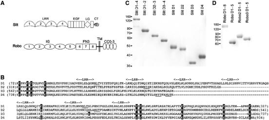
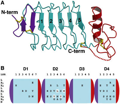
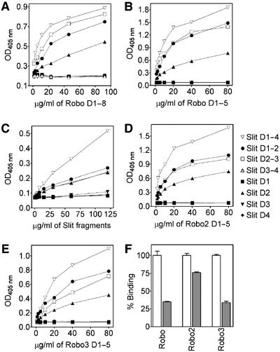
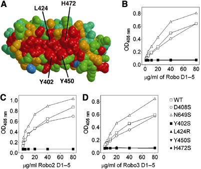
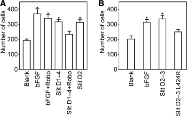
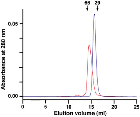
Recognition of the large secreted protein Slit by receptors of the Robo family provides fundamental signals in axon guidance and other developmental processes. In Drosophila, Slit-Robo signalling regulates midline crossing and the lateral position of longitudinal axon tracts. We report the functional dissection of Drosophila Slit, using structure analysis, site-directed mutagenesis and in vitro assays. The N-terminal region of Slit consists of a tandem array of four independently folded leucine-rich repeat (LRR) domains, connected by disulphide-tethered linkers. All three Drosophila Robos were found to compete for a single highly conserved site on the concave face of the second LRR domain of Slit. We also found that this domain is sufficient for biological activity in a chemotaxis assay. Other Slit activities may require Slit dimerisation mediated by the fourth LRR domain. Our results show that a small portion of Slit is able to induce Robo signalling and indicate that the distinct functions of Drosophila Robos are encoded in their divergent cytosolic domains.


Figures

Figure 1
Figure 1
Recombinant Slit and Robo constructs. (A) Domain organisation of Drosophila Slit and Robo: LRR, leucine-rich repeat; EGF, epidermal growth factor-like; LG, laminin G-like; CT, C-terminal cystine-knot; IG, immunoglobulin-like; FN3, fibronectin type 3-like; TM, transmembrane; CC0–3, conserved cytosolic motifs. (B) Sequence alignment of Drosophila Slit LRR domains D1–4. Cysteines are shaded black, putative N-linked glycosylation sites are underlined and the positions of LRR core motifs (LX1X2LX3LX4X5N) are indicated above the alignment. (C) Coomassie blue-stained reducing SDS–PAGE gel of recombinant His-myc-tagged Slit proteins used in this study. (D) Reducing SDS–PAGE gel of recombinant Robo proteins. Robo D1–8 is a fusion protein with a dimerising Fc-tag; all other Robo proteins have a C-terminal FLAG-tag. The positions of molecular mass markers are indicated on the left.
Figure 2
Figure 2
Structure of Slit LRR domains. (A) Cartoon drawing of the crystal structure of Slit D3. The N- and C-terminal caps are in violet and red, respectively, and LRRs 1–5 are in cyan. Disulphide bridges are in yellow. The N- and C-terminus are labelled. (B) Sequence conservation of the concave faces of D1–4. The domains are shown schematically in the orientation and colour scheme used in (A). The LRR consensus sequence is indicated on the left, running vertically from top to bottom. Within each domain, the LRRs are numbered horizontally and invariant residues at solvent-exposed positions X1–X5 are shown. Four residues in D2 that were mutated in this study are highlighted.
Figure 3
Figure 3
All three Drosophila Robos bind to Slit D2. (A) Binding of dimeric Fc-tagged Robo D1–8 to immobilised Slit fragments. (B) Binding of monomeric FLAG-tagged Robo D1–5 to immobilised Slit fragments. (C) Binding of His-myc-tagged Slit fragments to immobilised Robo D1–5. (D, E) Binding of FLAG-tagged Robo2 D1–5 (D) and Robo3 D1–5 (E) to immobilised Slit fragments. (F) Binding of FLAG-tagged Robos (50 μg/ml) to immobilised Slit D1–4 in the absence (open bars) and presence (grey bars) of a 10-fold excess of His-tagged Robo D1–5. The error bars indicate standard errors of the mean (n=3). Each experiment was carried out at least three times with similar results.
Figure 4
Figure 4
Conserved residues on the concave face of Slit D2 are important in Robo binding. (A) Sequence conservation mapped onto the concave face of a Slit D2 homology model (see text and Materials and methods). The colour scheme ranges from red (identity) to blue (no conservation). The orientation is similar to Figure 2A. Residues mutated in this study are labelled. (B–D) Binding of FLAG-tagged Robo D1–5 (B), Robo2 D1–5 (C) and Robo3 D1–5 (D) to immobilised Slit D2–3 proteins. Each experiment was carried out at least three times with similar results.
Figure 5
Figure 5
Slit D2 is sufficient to induce Robo-mediated HUVEC chemotaxis. (A) Comparison of the chemotactic activities of 0.4 nM basic fibroblast growth factor (bFGF; positive control) and 5 nM Slit proteins, as well as inhibition of Slit-induced chemotaxis by a 10-fold molar excess of Robo D1–5. (B) Comparison of chemotaxis induced by wild-type and mutant Slit D2–3 (5 nM). The number of migrated cells on the whole filter was determined by a person blinded to the experimental conditions. The bars indicate standard errors of the mean (n=5). An asterisk indicates significantly increased migration relative to unstimulated conditions (P<0.05, Student's t-test). Each experiment was carried out at least three times with similar results.
Figure 6
Figure 6
Dimerisation of Slit D4. Shown are gel filtration chromatograms of His-myc-tagged Slit D2 (blue; calculated monomer mass, 30.9 kDa; two N-linked carbohydrate chains) and Slit D4 (red; monomer mass, 27.5 kDa; one N-linked carbohydrate chain). Both proteins were injected at a concentration of 4 mg/ml. The elution volumes of two globular molecular mass standards are indicated by labelled arrows.

Similar articles

Cited by

References

    1. Araujo SJ, Tear G (2003) Axon guidance mechanisms and molecules: lessons from invertebrates. Nat Rev Neurosci 4: 910–922 - PubMed
    1. Barton WA, Liu BP, Tzvetkova D, Jeffrey PD, Fournier AE, Sah D, Cate R, Strittmatter SM, Nikolov DB (2003) Structure and axon outgrowth inhibitor binding of the Nogo-66 receptor and related proteins. EMBO J 22: 3291–3302 - PMC - PubMed
    1. Bashaw GJ, Kidd T, Murray D, Pawson T, Goodman CS (2000) Repulsive axon guidance: Abelson and Enabled play opposing roles downstream of the roundabout receptor. Cell 101: 703–715 - PubMed
    1. Battye R, Stevens A, Perry RL, Jacobs JR (2001) Repellent signaling by Slit requires the leucine-rich repeats. J Neurosci 21: 4290–4298 - PMC - PubMed
    1. Brose K, Bland KS, Wang KH, Arnott D, Henzel W, Goodman CS, Tessier-Lavigne M, Kidd T (1999) Slit proteins bind Robo receptors and have an evolutionarily conserved role in repulsive axon guidance. Cell 96: 795–806 - PubMed

Publication types

MeSH terms