. 2004 Nov 10;23(22):4371-83.
doi: 10.1038/sj.emboj.7600445. Epub 2004 Oct 21.

Evolving nature of the AP2 alpha-appendage hub during clathrin-coated vesicle endocytosis

Affiliations

Evolving nature of the AP2 alpha-appendage hub during clathrin-coated vesicle endocytosis

Gerrit J K Praefcke et al. EMBO J. .

Abstract

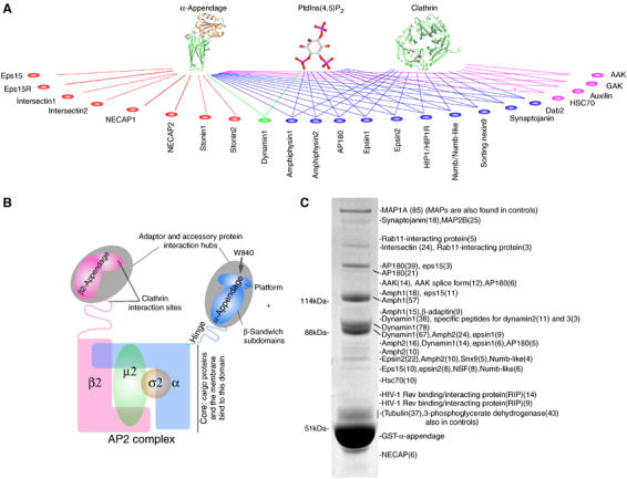
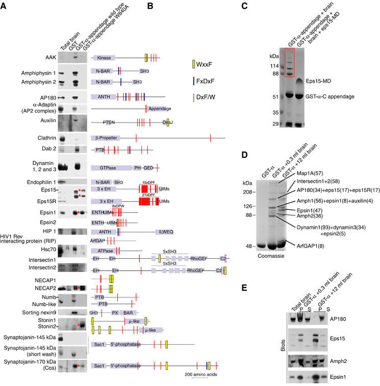
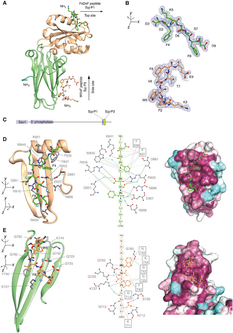
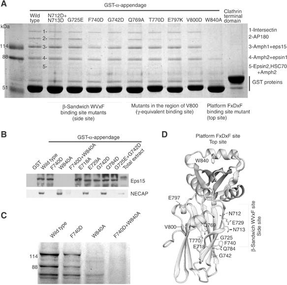
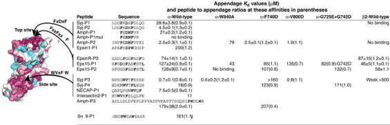
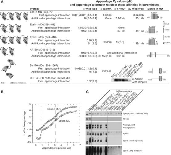
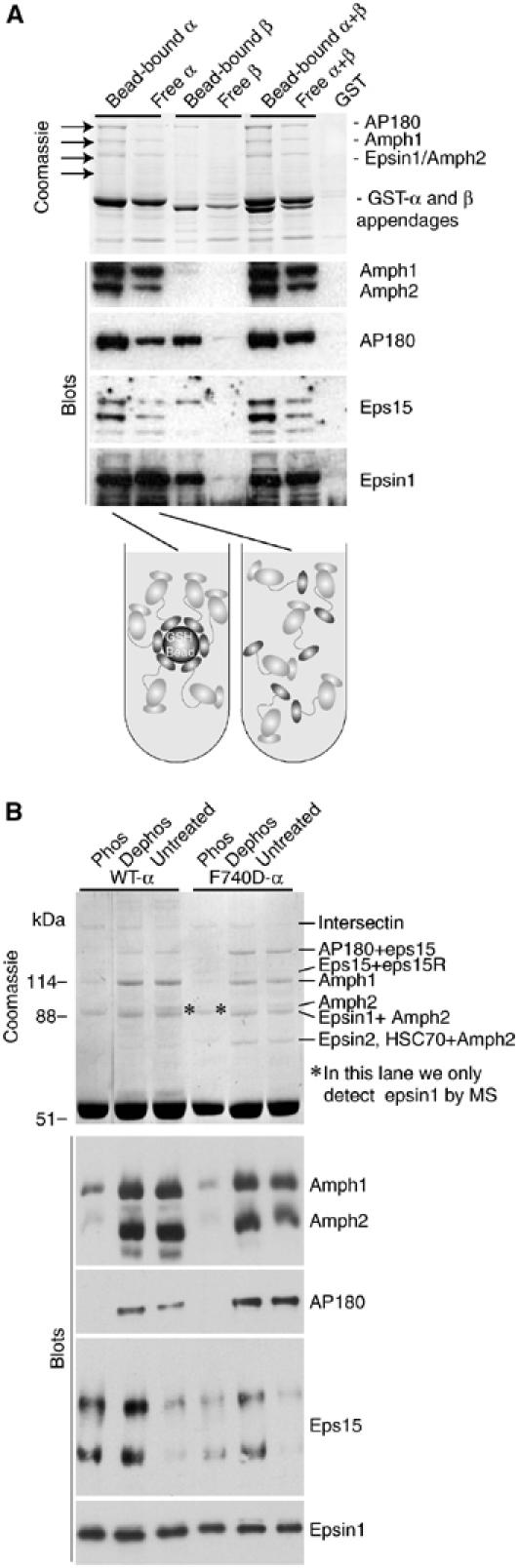
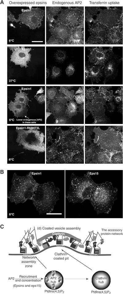
Clathrin-mediated endocytosis involves the assembly of a network of proteins that select cargo, modify membrane shape and drive invagination, vesicle scission and uncoating. This network is initially assembled around adaptor protein (AP) appendage domains, which are protein interaction hubs. Using crystallography, we show that FxDxF and WVxF peptide motifs from synaptojanin bind to distinct subdomains on alpha-appendages, called 'top' and 'side' sites. Appendages use both these sites to interact with their binding partners in vitro and in vivo. Occupation of both sites simultaneously results in high-affinity reversible interactions with lone appendages (e.g. eps15 and epsin1). Proteins with multiple copies of only one type of motif bind multiple appendages and so will aid adaptor clustering. These clustered alpha(appendage)-hubs have altered properties where they can sample many different binding partners, which in turn can interact with each other and indirectly with clathrin. In the final coated vesicle, most appendage binding partners are absent and thus the functional status of the appendage domain as an interaction hub is temporal and transitory giving directionality to vesicle assembly.


Figures

Figure 1
Figure 1
Proteomic analysis of AP2 α-appendage interactions. (A) Scheme showing all α-appendage binding partners, highlighting those that also interact with other interaction hubs in clathrin-mediated endocytosis. Colours separate interaction groups. (B) Scheme of an AP2 adaptor complex with its two appendage domains. The α-appendage is the main accessory protein interaction hub. (C) Protein from brain extracts bound to the α-appendage and analysed by tandem mass spectrometry. Numbers in brackets indicate the number of peptides sequenced to confirm the identity of the band. More details of interacting proteins are given in Supplementary Table 1.
Figure 2
Figure 2
Protein interactions with AP2 α-appendage domain. (A) Protein from brain extracts bound to the α-appendage and the α mutant W840A. The red stars indicate proteins that are not displaced completely by the α-W840A mutation implying an additional interaction mode. 100 μg of GST fusion protein were used in 10 mg of rat brain extract and 6 μg of fusion protein were loaded for blotting. Given the number of interacting partners for the α-appendage, this excess of GST fusion protein allows us to sample all possible interacting partners without saturating the fusion proteins with higher affinity ligands. Bead-bound proteins were washed for 10 min. (B) Domains of α-appendage ligands showing the predicted α-binding motifs clustered in many cases into MDs. These are not all functional motifs but some have been tested in Figures 4 and 5. Note that for eps15 a polyclonal antibody was used and that only the upper two forms detected by the antibody bind to the appendages. As there is very little of the 170 kDa form of synaptojanin in the brain, we probed the extract from a 90 mm dish of COS cells. We have used a pan-dynamin antibody and by mass spectrometry we found dynamins I, II and III bound in the α-appendage complex. Intersectins 1 and 2, epsin2, NECAP and sorting nexin9 were identified as α-appendage ligands in mass spectrometry of these samples (see Figure 1C). (C) A Coomassie-stained gel showing a five-fold molar excess of eps15 MD in brain extract competes off all ligands from the α-appendage. (D) GST-α-appendage interactions in 0.3 and 12 ml of brain extract. The major proteins detected by Coomassie staining that change intensity have been identified by LC-MS/MS and the number of peptides is in parentheses. (E) Blots of samples in (D). AAK: adaptor-associated kinase; Amph: amphiphysin; Dab2: disabled protein2; HIP1: Huntingtin interacting protein1; UIMs: ubiquitin interacting motifs; PTB: phosphotyrosine binding domain; BAR: Bin/amphiphysin/Rvs homology domain; PX: phox homology domain.
Figure 3
Figure 3
α-Adaptin appendage bound to WVxF and FxDxF motifs from synaptojanin. (A) Ribbon diagram showing bound peptides. The β-sandwich subdomain is coloured green and the platform subdomain is gold. The dark green residues at the N-terminus are from the vector. Syj-P3 and Syj-P1 bound to the α-appendage have been deposited with the Protein Data Bank (PDB ids: 1w80 and r1w80sf). (B) Density profiles for the peptides. (C) Scheme of the position of P1 and P3 peptides in synaptojanin170. (D, E) Details of peptide binding sites showing critical α-appendage residues and coordinated waters (blue) involved in binding. Peptides displayed as a linear chain are in the centre panel showing hydrogen bonding potential (dashed green lines) and hydrophobic (grey lines) interactions. The most crucial peptide residues are in bold italics and the residues for which there is little or no density are dotted. On the right are surface representations around the peptide binding sites, coloured according to sequence conservation (maroon (well conserved) through white to light blue (not conserved)). Binding pockets are more easily visible in this representation.
Figure 4
Figure 4
Top and side interaction surfaces on the α-appendage. (A) Coomassie analysis of ligand binding to α-appendage mutants. The results of a mass spectrometry analysis of the major Coomassie bands 1–5 are given on the right. (B) We found that NECAP does not bind to α-F740D and thus we probed some mutants around this site along with the top site mutant W840A. (C) Coomassie analysis of mutants with less stringent washing than in (A) leaving visible Coomassie bands in α-W840A that are absent in the double top and side site mutant. (D) Positions of our α-appendage mutants.
Figure 5
Figure 5
Affinities of peptides measured by ITC. Dissociation constants for the interactions of peptides and α-appendages as determined by ITC at 10°C. Where we have more than one determination, we give the average±range. The stoichiometries are in parentheses. The Amph-P3 peptide is from amphiphysin2 and contains two adaptor binding sequences, one for the platform and the other for the β-sandwich subdomain, neither of which have been previously shown. A stoichiometry of 2:1 means one peptide cannot stretch between the two appendage sites. The surface conservation to the left is colour coded: burgandy for high conservation through white to blue/jade for residues not conserved. Syj: synaptojanin; Amph: amphiphysin; Snx9: sorting nexin9. Further ITC parameters are found in Supplementary Table 3.
Figure 6
Figure 6
Affinities of proteins measured by ITC. (A) Dissociation constants and other binding parameters for the interactions of MD with α-appendages at 10°C measured by ITC. Schematic diagrams of the appendage MD interactions are on the left and schematics of the MD constructs used are on the right; numbers refer to amino acids. Inset: pull-downs from brain extract with some of the same MDs showing that affinities measured by ITC match the amounts of AP2 bound. Further ITC parameters are found in Supplementary Table 4. (B) Typical ITC data showing separation of high- and medium-affinity binding sites in eps15-MD and epsin1-MD+. (C) Peptide competition for ligand binding from brain extract to GST-α-appendage mutants. All peptides were used at 100 μM. The sequence of LLDLD peptides from amphiphysin1 is KEETLLDLDFDPF. This peptide and Amph-P3 bind to clathrin terminal domains. Other peptide sequences are found in Figure 5. As in Figure 2A, COS cell extract was used to monitor the interaction of synaptojanin170. Beads were washed for at least 10 min.
Figure 7
Figure 7
Influence of clustering and phosphorylation on α-appendage interactions. (A) Free appendages versus clustered appendages have different ligand preferences. As an approximation to free appendages, we have incubated GST-appendages in brain extract and subsequently captured these by pouring the extract over a filter with GSH beads on top. This was compared with GST-appendages previously bound to beads and then incubated in brain extract. The Coomassie gel shows the major changes taking place and some proteins have been blotted. The identities of the Coomassie bands have not been determined by mass spectrometry. (B) α-Appendages incubated in phosphorylated, dephosphorylated and untreated brain extracts change their ligand preferences. The identities of dominant proteins by LC-MS/MS are given beside the Coomassie gel.
Figure 8
Figure 8
Adaptor protein-network assembly-zones. (A) Overexpression of epsin1 in COS cells leads to a temperature-dependent accumulation of puncta. This results in a redistribution of endogenous AP2 into these same puncta. Their presence is absent in cells expressing low levels of endogenous AP2 (50 out of 50 cells). The epsin1 lipid binding mutant R63L+H73L, which we previously showed inhibits transferrin at 37°C, does not form these puncta at 6°C and indeed AP2 becomes aggregated in the cytosol, but does not colocalise with other endocytic markers (not shown). Based on these experiments and the temperature-dependent appearance and our previous observations of colocalisation with eps15 and dynamin, we call these puncta ‘adaptor protein-network assembly-zones'. Scale bar, 20 μm. (B) Colocalisation of endogenous eps15 (detected with Ra15) with epsin1 in puncta. Scale bar, 20 μm. (C) Model of clathrin-coated pit assembly illustrating the adaptor protein-network assembly-zones at the edges of the pit. In these zones, proteins like epsins and eps15 are capturing more adaptors, so the vesicles are filled with cargo and shaping the membrane in preparation for clathrin assembly. Proteins with α-appendage side site interaction motifs will be able to bind simultaneously to the α-appendage along with epsins and eps15, which cluster primarily by using the top site. Thus, synaptojanin may well be recruited at this point and with its lipid phosphatase activity it will be able to destabilise the membrane attachments unless there is prior clathrin polymerisation. Amphiphysins are also found here and recruit dynamin to the edges of coated pits in preparation for the vesicle scission process. Assembly-zones may be too small and too unstable to visualise at 37°C by microscopy. A stable network will naturally progress into a coated pit due to the presence of clathrin-polymerising molecules like amphiphysin, epsins and AP180. This means that the network self-destructs as clathrin is polymerised and thus the network does not grow indefinitely. The initial AP2 recruitment to membranes is likely aided by high-affinity single appendage interactions like those for epsin1 and eps15. The affinity of both AP2 and epsin1 for membranes and cargo will help stabilise the membrane interactions.

Similar articles

Cited by

References

    1. Brett TJ, Traub LM, Fremont DH (2002) Accessory protein recruitment motifs in clathrin-mediated endocytosis. Structure (Camb) 10: 797–809 - PubMed
    1. Brodsky FM, Chen CY, Knuehl C, Towler MC, Wakeham DE (2001) Biological basket weaving: formation and function of clathrin-coated vesicles. Annu Rev Cell Dev Biol 17: 517–568 - PubMed
    1. Chen H, Slepnev VI, Di Fiore PP, De Camilli P (1999) The interaction of epsin and Eps15 with the clathrin adaptor AP-2 is inhibited by mitotic phosphorylation and enhanced by stimulation-dependent dephosphorylation in nerve terminals. J Biol Chem 274: 3257–3260 - PubMed
    1. Collins BM, Praefcke GJ, Robinson MS, Owen DJ (2003) Structural basis for binding of accessory proteins by the appendage domain of GGAs. Nat Struct Biol 10: 607–613 - PubMed
    1. Conner SD, Schmid SL (2003) Regulated portals of entry into the cell. Nature 422: 37–44 - PubMed

Publication types

MeSH terms