. 2004 Dec 15;561(Pt 3):721-34.
doi: 10.1113/jphysiol.2004.077339. Epub 2004 Oct 21.

Slc26a6: a cardiac chloride-hydroxyl exchanger and predominant chloride-bicarbonate exchanger of the mouse heart

Affiliations

Slc26a6: a cardiac chloride-hydroxyl exchanger and predominant chloride-bicarbonate exchanger of the mouse heart

Bernardo V Alvarez et al. J Physiol. .

Abstract

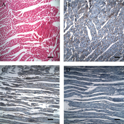
Bicarbonate facilitate more than 50% of pH recovery in the acidotic myocardium, and have roles in cardiac hypertrophy and steady-state pH regulation. To determine which bicarbonate transporters are responsible for this activity, we measured the expression levels of all known HCO3(-)-anion exchange proteins in mouse heart, by quantitative real time RT-PCR. Bicarbonate-anion exchangers are members of either the SLC4A or the SLC26A gene families. In neonatal and adult myocardium, AE1 (Slc4a1), AE2 (Slc4a2), AE3 (Slc4a3) (AE3fl and AE3c variants), Slc26a3 and Slc26a6 were expressed. Adult hearts expressed Slc26a3 and Slc4a1-3 mRNAs at similar levels, while Slc26a6 mRNA was about seven-fold higher than AE3, which was more abundant than any other. Immunohistochemistry revealed that Slc26a6 and AE3 are present in the plasma membrane of ventricular myocytes. Slc26a6 expression levels were higher in ventricle than atrium, whereas AE3 was detected only in ventricle. Cl(-)-HCO(3)(-) and Cl(-)-OH(-) exchange activity of SLC26A6 and AE3 were investigated in transfected HEK293 cells, using intracellular fluorescence measurements of 2',7'-bis (2-carboxyethyl)-5(6)-carboxyfluorescein (BCECF), to monitor intracellular pH (pH(i)). Rates of pH(i) change were measured under HCO3(-)-containing (Cl(-)-HCO(3)(-)) or nominally HCO3(-)-free (Cl(-)-OH(-)) conditions. HCO3(-) fluxes were similar for cells expressing AE3fl, SLC26A6 or Slc26a3, suggesting that they have similar transport activity. However, only SLC26A6 and Slc26a3 functioned as Cl(-)-OH(-) exchangers. Activation of alpha-adrenergic receptors, which stimulates protein kinase C, inhibited SLC26A6 Cl(-)-HCO(3)(-) exchange activity. We conclude that Slc26a6 is the predominant Cl(-)-HCO(3)(-) and Cl(-)-OH(-) exchanger of the myocardium and that Slc26a6 is negatively regulated upon alpha-adrenergic stimulation.


Figures

Figure 1
Figure 1. Expression of Slc26 transcripts in adult mouse heart atria and ventricles
mRNA expression was compared in samples from atria and ventricles of adult mice using real time quantitative RT-PCR. Data are corrected for individual variation using glyceraldehyde-3-phosphate dehydrogenase (GAPDH) standard curves and results are expressed as cycle threshold. The cycle threshold is dependent on the quantity of the target molecule in the sample. The higher the cycle threshold value, the lower the expression of the transcript. Dashed line marked limit of expressed transcripts. Cycle threshold values of 32 or higher means no expression of the transcript. Error bars represent s.e.m. (n = 3 separate adult mouse hearts supplying atrial and ventricular samples).
Figure 2
Figure 2. Expression of anion exchanger transcripts in mouse heart and quantification by real time quantitative RT-PCR
A, RT-PCR analysis of various anion exchangers in adult mouse ventricle. First strand cDNA was produced using First Strand RT-PCR Kit. Amplicons were analysed on a 1% agarose–ethidium bromide gel. Lane 1, AE1; lane 2, AE2; lane 3, AE3c; lane 4, AE3fl; lane 5, Slc26a6; lane 6, Slc26a3. B and C, mRNA expression was compared from ventricles of adult (filled bars) or neonatal (grey bars) mice using real time quantitative RT-PCR. Data are corrected for individual variation using plasmid DNA standards and results are expressed as copy number (logarithmic scale). Two different pairs of forward and reverse primers (1 and 2), which amplified different regions of Slc26a6 cDNA, and which had slightly different amplicon size and Tm (see Table 1), were used. *Significant difference from the expression level of Slc26a6 (primer set 1) in adult ventricle, with P < 0.05, one-way ANOVA. #P < 0.05 (paired t test) compared to Slc26a6 (primer set 2) in adult heart samples. γNeonatal samples different from the expression level of Slc26a6 (primer set 1) with P < 0.05, one-way ANOVA. Error bars represent s.e.m. (n = 4 separate hearts for adults and 3 for neonates).
Figure 3
Figure 3. Expression of Slc26a6 and AE3 proteins in adult mouse heart atria and ventricles, and expression of Slc26a6 protein in isolated adult mouse cardiomyocytes
A and B, heart membranes were isolated from adult mouse atria and ventricles. Membrane protein (50 μg) was subjected to SDS-PAGE analysis, transferred to PVDF membrane, and probed with specific anti-SLC26A6 (A), or anti-AE3 (B) antibodies. C and D, lysates of HEK293 cells (20 μg protein) transfected with either empty vector (lane 1), or transfected with human SLC26A6 cDNAs (lane 2), or lysates of freshly isolated mouse cardiomyocytes (50 μg, lane 3), were prepared. Samples were analysed by SDS-PAGE, transferred to PVDF membranes, and probed with anti-SLC26A6 antibody (C) or serum from nonimmune rabbits (D). Filled and open arrows indicate positions of the SLC26A6–Slc26a6 and AE3 proteins, respectively.
Figure 4
Figure 4. Immunocytochemistry of Slc26a6 and AE3 proteins in adult mouse cardiac ventricle
A, haematoxylin–eosin staining of ventricular muscle sections. Immunostaining of Slc26a6 protein (B) and AE3 protein (C) was detected on the surface of cardiomyocytes, in ventricular muscle sections. No immunoreactivity was seen on cardiac muscle sections processed with pre-immune rabbit serum (D). Scale bar represents 25 μm.
Figure 5
Figure 5. Chloride–base exchange activity of cardiac anion exchange proteins
A, HEK293 cells were transfected with SLC26A6 cDNA. Two days after transfection, cells were loaded with pH-sensitive dye, BCECF. Cells were perfused with either Cl-free (filled bar), or Cl-containing (open bar) buffer. Dark and light traces show experiments performed using bicarbonate- and Hepes-buffered Tyrode solutions, respectively. B, mean values of transport rates expressed as mmol l−1 min−1, of HEK293 cell transiently transfected with SLC26A6 cDNA. Cells expressing human SLC26A6 were exposed to DIDS (1 mm). Filled and grey bars represent experiments performed in bicarbonate- and Hepes-buffered Tyrode solutions, respectively. *P < 0.05, paired t test. C, Cl–HCO3 (bicarbonate) and Cl–OH (Hepes) exchange activity was measured in HEK293 cells transiently transfected with rat AE3fl (rAE3fl), human SLC26A6 (hSLC26A6), mouse Slc26a3 (mSlc26a3) cDNAs, or empty vector (pCDNA3). Transport rates are expressed as flux (mmol l−1 min−1); (number of experiments). *P < 0.05 compared to pCDNA3, γP < 0.05 compared to hSLC26A6; ζP < 0.05 compared to rAE3fl; one-way ANOVA.
Figure 6
Figure 6. Negative regulation of Slc26a6 Cl–HCO3 exchange activity upon α1a-adrenergic receptor stimulation
A, expression of haemaglutinin epitope-tagged α1a receptors (α1a-HA), in HEK293 cells. Lysates of HEK293 cells (20 μg protein) transfected with either empty vector (lane 1), or transfected with α1a-HA cDNA (lane 2), were prepared. Samples were analysed by SDS-PAGE, transferred to PVDF membranes, and probed with anti-HA antibody. Open arrows indicate position of different glycosylated forms of the α1a receptor. B, mean values of Cl–HCO3 exchange activity relative to SLC26A6 transport activity, of HEK293 cell transiently transfected with SLC26A6, or cotransfected with SLC26A6 and α1a-HA, cDNAs, as indicated at the bottom. Cells expressing human SLC26A6 and α1a receptors were incubated with phenylephrine (PE, 10 μm), or with PMA-phorbol esters (200 nm) to stimulate protein kinase C (PKC). Chelerythrine (CHE, 10 μm) was used to block PKC activity in cells expressing SLC26A6. *P < 0.05, paired t test.

References

    1. Alvarez BV, Fujinaga J, Casey JR. Molecular basis for angiotensin II-induced increase of chloride/bicarbonate exchange in the myocardium. Circ Res. 2001;89:1246–1253. - PubMed
    1. Boron WF, De Weer P. Intracellular pH transients in squid giant axons caused by CO2, NH3, and metabolic inhibitors. J Gen Physiol. 1976;67:91–112. - PMC - PubMed
    1. Bouchard RA, Clark RB, Giles WR. Regulation of unloaded cell shortening by sarcolemmal sodium–calcium exchange in isolated rat ventricular myocytes. J Physiol. 1993;469:583–599. - PMC - PubMed
    1. Camilion de Hurtado MC, Alvarez BV, Ennis IL, Cingolani HE. Stimulation of myocardial Na+-independent Cl−-HCO3− exchanger by angiotensin II is mediated by endogenous endothelin. Circ Res. 2000;86:622–627. - PubMed
    1. Camilion de Hurtado MC, Alvarez BV, Perez NG, Ennis IL, Cingolani HE. Angiotensin II activates Na+-independent Cl−-HCO3− exchange in ventricular myocardium. Circ Res. 1998;82:473–481. - PubMed

Publication types

LinkOut - more resources