. 2004 Nov 23;101(47):16442-7.
doi: 10.1073/pnas.0405809101. Epub 2004 Oct 21.

3-hydroxy-3-methylglutaryl-CoA synthase intermediate complex observed in "real-time"

Affiliations

3-hydroxy-3-methylglutaryl-CoA synthase intermediate complex observed in "real-time"

Michael J Theisen et al. Proc Natl Acad Sci U S A. .

Abstract

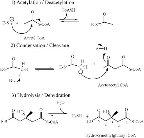
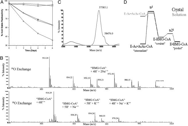
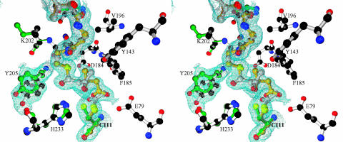
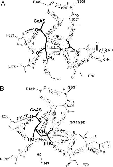
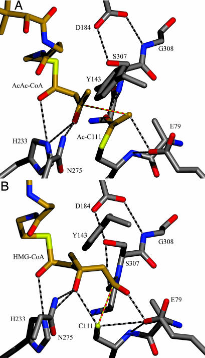
The formation of carbon-carbon bonds via an acyl-enzyme intermediate plays a central role in fatty acid, polyketide, and isoprenoid biosynthesis. Uniquely among condensing enzymes, 3-hydroxy-3-methylglutaryl (HMG)-CoA synthase (HMGS) catalyzes the formation of a carbon-carbon bond by activating the methyl group of an acetylated cysteine. This reaction is essential in Gram-positive bacteria, and represents the first committed step in human cholesterol biosynthesis. Reaction kinetics, isotope exchange, and mass spectroscopy suggest surprisingly that HMGS is able to catalyze the "backwards" reaction in solution, where HMG-CoA is cleaved to form acetoacetyl-CoA (AcAc-CoA) and acetate. Here, we trap a complex of acetylated HMGS from Staphylococcus aureus and bound acetoacetyl-CoA by cryo-cooling enzyme crystals at three different times during the course of its back-reaction with its physiological product (HMG-CoA). This nonphysiological "backwards" reaction is used to understand the details of the physiological reaction with regards to individual residues involved in catalysis and substrate/product binding. The structures suggest that an active-site glutamic acid (Glu-79) acts as a general base both in the condensation between acetoacetyl-CoA and the acetylated enzyme, and the hydrolytic release of HMG-CoA from the enzyme. The ability to trap this enzyme-intermediate complex may suggest a role for protein dynamics and the interplay between protomers during the normal course of catalysis.


Figures

Scheme 1.
Scheme 1.
Reactions catalyzed by HMGS.
Fig. 1.
Fig. 1.
Monitoring different aspects of the “backwards” reaction catalyzed by HMGS. (A) HMGS catalyzes the cleavage of HMG–CoA; solid triangles and open circles represent wild-type avian and S. aureus enzymes, respectively; open squares and open diamonds represent the avian C129S mutated enzyme and BSA, respectively; and open triangles represent a 2× concentration of wild-type avian enzyme. (B) Mass spectra of HMG–CoA incubated for 30 min with 16O(Upper) or 18O(Lower) water in the presence of wild-type enzyme. This example shows that nearly complete exchange of the carboxylate oxygens has occurred. (C) Mass spectra of the E95A mutated enzyme after a 5-day incubation with HMG–CoA shows that this enzyme is capable of forming a covalent intermediate. (D) Energy profile for the latter half of the reaction pathway in the crystal and in solution.
Fig. 2.
Fig. 2.
Stereo diagram of the electron density map (2FoFc) surrounding the CoA moiety, Cys-111, and Tyr-205 from one of the active sites in the day 31 crystal superimposed on the final atomic model. Residues modeled with green bonds correspond specifically to the apo enzyme, residues modeled with gold bonds correspond to the HMG–CoA complex, residues modeled with silver bonds correspond to the AcAc–CoA complex, and residues modeled with white bonds correspond to common conformations. The position of the sulfur of Cys-111 that is nearest to the observer corresponds to the position when HMG–CoA is bound, whereas the position to the left corresponds to the apo enzyme conformation. The apo enzyme conformation is sterically incompatible with the binding of HMG–CoA. Note how the modeled alternative conformation of Tyr-205 helps to explain the observed electron density.
Fig. 3.
Fig. 3.
Schematic representation of the contacts in the active site with AcAc–CoA (A) and the acetylcysteine and HMG–CoA (B). The wide dashed lines indicate close contacts that violate van der Waals distance constraints, and the narrow dashed lines indicate potential hydrogen bonds. The average distances are given in angstroms (10–10 m) based on either 11 or 7 structures, and the numbers in parentheses are the standard deviations of the distance multiplied by 100.
Fig. 4.
Fig. 4.
Models of the AcAc–CoA-acetylated enzyme complex (A) and the HMG–CoA enzyme complex (B). The atoms are colored according to their CPK atom type, and the bonds are shown in either white (for protein) or gold (for the acetyl moiety and the CoA molecules). Yellow-and-red dashed bonds tie reacting atoms together.

Comment in

Similar articles

Cited by

References

    1. Greenspan, M. D., Yudkovitz, J. B., Lo, C. Y., Chen, J. S., Alberts, A. W., Hunt, V. M., Chang, M. N., Yang, S. S., Thompson, K. L., Chiang, Y. C., et al. (1987) Proc. Natl. Acad. Sci. USA 84, 7488–7492. - PMC - PubMed
    1. Teo, K. K. & Burton, J. R. (2002) Drugs 62, 1707–1715. - PubMed
    1. Wilding, E. I., Brown, J. R., Bryant, A. P., Chalker, A. F., Holmes, D. J., Ingraham, K. A., Iordanescu, S., So, C. Y., Rosenberg, M. & Gwynn, M. N. (2000) J. Bacteriol. 182, 4319–4327. - PMC - PubMed
    1. Campobasso, N., Patel, M., Wilding, E. I., Kallender, H., Rosenberg, M. & Gwynn, M. (2004) J. Biol. Chem. 279, 44883–44888. - PubMed
    1. Edwards, D. J., Marquez, B. L., Nogle, L. M., McPhail, K., Goeger, D. E., Roberts, M. A. & Gerwick, W. H. (2004) Chem. Biol. 11, 817–833. - PubMed

Publication types

LinkOut - more resources