

A novel method of identifying genetic mutations using an electrochemical DNA array
- PMID: 15498924
- PMCID: PMC524315
- DOI: 10.1093/nar/gnh141
A novel method of identifying genetic mutations using an electrochemical DNA array
Erratum in
- Nucleic Acids Res. 2004 Nov 24;32(20):6152
Abstract
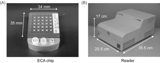
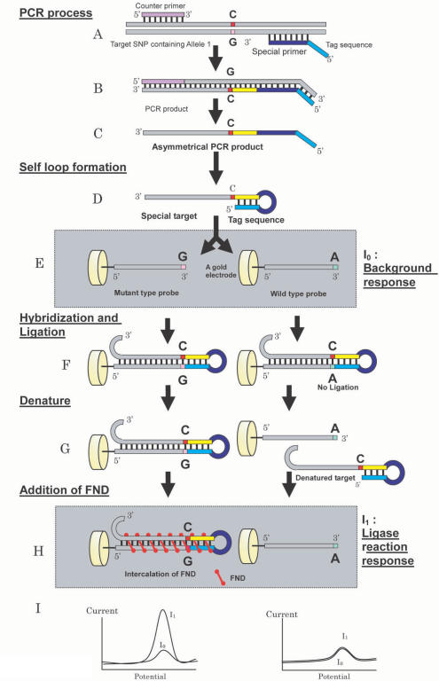
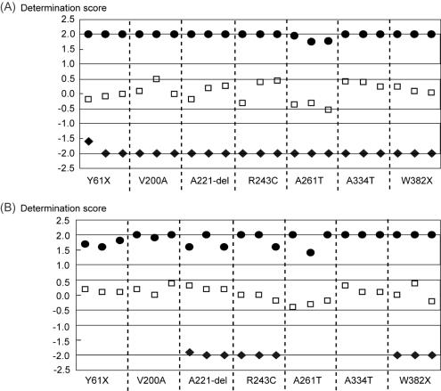
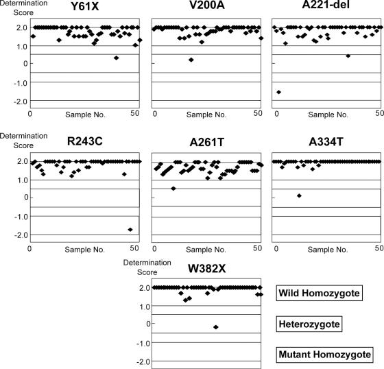
We describe the development of a new type of DNA array chip that utilizes electrochemical reactions and a novel method of simultaneously identifying multiple genetic mutations on an array chip. The electrochemical array (ECA) uses a threading intercalator specific to double-stranded nucleotides, ferrocenylnaphthalene diimide (FND), as the indicator. ECA does not require target labeling, and the equipment is simple, durable and less expensive. The simultaneous multiple mutation detection (SMMD) system using an ECA chip and FND utilizes an enzyme to simultaneously distinguish several genetic mutations such as single nucleotide polymorphism (SNP), insertion, deletion, translocation and short tandem repeat. We examined this SMMD system using an ECA chip, by detecting seven different mutations on the lipoprotein lipase (LPL) gene for 50 patients in a blind test. It turned out that all the results obtained were concordant with the sequencing results, demonstrating that this system is a powerful tool for clinical applications.
Figures

Similar articles
-
Genotyping of the human lipoprotein lipase gene by ferrocenylnaphthalene diimide-based electrochemical hybridization assay.Anal Sci. 2005 Dec;21(12):1437-41. doi: 10.2116/analsci.21.1437. Anal Sci. 2005. PMID: 16379382
-
Direct detection of single nucleotide polymorphism (SNP) with genomic DNA by the ferrocenylnaphthalene diimide-based electrochemical hybridization assay (FND-EHA).Anal Sci. 2003 Jan;19(1):79-83. doi: 10.2116/analsci.19.79. Anal Sci. 2003. PMID: 12558028
-
Electrochemical detection of telomeric quadruplex DNA using ferrocenyl naphthalene diimide.Nucleic Acids Symp Ser (Oxf). 2005;(49):237-8. doi: 10.1093/nass/49.1.237. Nucleic Acids Symp Ser (Oxf). 2005. PMID: 17150721
-
Electrochemical DNA detection using supramolecular interactions.Anal Sci. 2012;28(7):643-9. doi: 10.2116/analsci.28.643. Anal Sci. 2012. PMID: 22790364 Review.
-
Proofreading genotyping assays and electrochemical detection of SNPs.Hum Mutat. 2004 May;23(5):420-5. doi: 10.1002/humu.20034. Hum Mutat. 2004. PMID: 15108272 Review.
Cited by
-
Advances in nanomaterials and their applications in point of care (POC) devices for the diagnosis of infectious diseases.Biotechnol Adv. 2016 Dec;34(8):1275-1288. doi: 10.1016/j.biotechadv.2016.09.003. Epub 2016 Sep 26. Biotechnol Adv. 2016. PMID: 27686397 Free PMC article. Review.
-
DNA single-base mismatch study with an electrochemical enzymatic genosensor.Biosens Bioelectron. 2007 Mar 15;22(8):1642-50. doi: 10.1016/j.bios.2006.07.015. Epub 2006 Sep 1. Biosens Bioelectron. 2007. PMID: 16950611 Free PMC article.
-
DNA diagnostics: nanotechnology-enhanced electrochemical detection of nucleic acids.Pediatr Res. 2010 May;67(5):458-68. doi: 10.1203/PDR.0b013e3181d361c3. Pediatr Res. 2010. PMID: 20075759 Free PMC article. Review.
-
Impedimetric Sensing of Factor V Leiden Mutation by Zip Nucleic Acid Probe and Electrochemical Array.Biosensors (Basel). 2020 Sep 7;10(9):116. doi: 10.3390/bios10090116. Biosensors (Basel). 2020. PMID: 32906640 Free PMC article.
-
An electrochemical sensor for single nucleotide polymorphism detection in serum based on a triple-stem DNA probe.J Am Chem Soc. 2009 Oct 28;131(42):15311-6. doi: 10.1021/ja905068s. J Am Chem Soc. 2009. PMID: 19807078 Free PMC article.
References
Publication types
MeSH terms
Substances
LinkOut - more resources
Full Text Sources
Other Literature Sources
