. 2005 Jan;25(1):102-8.
doi: 10.1161/01.ATV.0000148547.70187.89. Epub 2004 Oct 21.

Uterine spiral artery remodeling involves endothelial apoptosis induced by extravillous trophoblasts through Fas/FasL interactions

Affiliations

Uterine spiral artery remodeling involves endothelial apoptosis induced by extravillous trophoblasts through Fas/FasL interactions

Sandra V Ashton et al. Arterioscler Thromb Vasc Biol. 2005 Jan.

Abstract

Objective: Invasion of uterine spiral arteries by extravillous trophoblasts in the first trimester of pregnancy results in loss of endothelial and musculoelastic layers. This remodeling is crucial for an adequate blood supply to the fetus with a failure to remodel implicated in the etiology of the hypertensive disorder preeclampsia. The mechanism by which trophoblasts induce this key process is unknown. This study gives the first insights into the potential mechanisms involved.

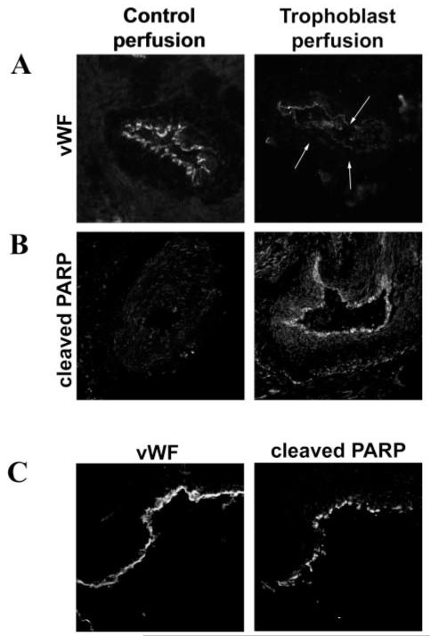
Methods and results: Spiral arteries were dissected from nonplacental bed biopsies obtained at Caesarean section, and a novel model was used to mimic in vivo events. Arteries were cultured with trophoblasts in the lumen, and apoptotic changes in the endothelial layer were detected after 20 hours, leading to loss of endothelium by 96 hours. In vitro, coculture experiments showed that trophoblasts stimulated apoptosis of primary decidual endothelial cells and an endothelial cell line. This was blocked by caspase inhibition and NOK2, a FasL blocking antibody. NOK2 also abrogated trophoblast-induced endothelial apoptosis in the vessel model.

Conclusions: Extravillous trophoblast induction of endothelial apoptosis is a possible mechanism by which the endothelium is removed, and vascular remodeling may occur in uterine spiral arteries. Fas/FasL interactions have an important role in trophoblast-induced endothelial apoptosis.


Figures

Figure 1
Figure 1. Trophoblasts induce spiral artery EC loss through apoptosis.
Unmodified, nonplacental bed uterine spiral arteries were perfused with CellTracker-fluorescently labeled SGHPL-4 cells or primary first-trimester cytotrophoblasts or media (control). Vessels were incubated for periods of 20 to 96 hours before sectioning. A, Expression of vWF indicating the presence of endothelium in the control perfusion and absence of endothelium in vessels cultured with primary trophoblasts for 96 hours (the position of the lumen is indicated by the arrows). B, Expression of p85-cleaved PARP in the endothelium of trophoblast-perfused vessels, incubated for 20 hours. C, Colocalization of cleaved PARP with vWF staining in consecutive sections from vessels cultured with SGHPL-4 cells, confirming apoptosis in the endothelium. Original magnification ×150.
Figure 2
Figure 2. Coculture with trophoblasts in vitro leads to EC apoptotic morphology.
A, SGHPL-4 cells were cocultured with fluorescently labeled SGHEC-7 cells or decidual ECs shown at 0 hours as a fluorescent and phase-contrast image. Apoptotic morphology could be detected in ECs over time, evidenced by cytoplasmic retraction and a phase-bright appearance, and membrane blebbing and blistering (indicated by arrows). Apoptosis in an individual EC is shown to illustrate the morphological changes occurring over time. B, Apoptosis of SGHEC-7 and primary decidual ECs cocultured with trophoblasts was monitored by time-lapse microscopy. Cells were scored by the time point at which clear apoptotic morphology was observed. At least 3 separate experiments were performed, and 40 cells were assessed per well in each experiment. Pooled mean data are shown. Error bars have been omitted for clarity because of the number of time points. Apoptotic morphological changes could be detected throughout the experiment; however, the difference between EC apoptosis in the presence of trophoblasts compared with ECs alone reached statistical significance after 10 hours (P=0.04; n=9) for SGHEC-7 cells and after 21 hours (P=0.04; n=5) for decidual ECs. zVAD-fmk was added at the same time as trophoblasts.
Figure 3
Figure 3. Expression of cleaved PARP by ECs cocultured with trophoblasts.
SGHEC-7 cells were cultured for 24 hours or 60 hours with SGHPL-4 trophoblast cells or SGHEC-7 (to control for cell number) in tissue culture inserts before lysis of the ECs. Western blot analysis with rabbit polyclonal anti-human cleaved PARP showed a band at 85 kDa. Actin loading control is shown with a band at 42 kDa. Lane 1, 24-hour endothelial culture; lane 2, 24-hour endothelial+trophoblast coculture; lane 3, 60-hour endothelial culture; lane 4, 60-hour endothelial+trophoblast coculture.
Figure 4
Figure 4. Fas expression by spiral arteries and ECs.
A, Immunohistochemical analysis of Fas expression in spiral arteries. Sections were labeled with anti-Fas, anti-vWF, or control immunoglobulin in consecutive sections. Original magnification ×150. B, Western blot analysis of Fas expression (45 KDa) after immunoprecipitation with polyclonal anti-Fas and detection with monoclonal anti-Fas. Lane 1, Decidual ECs; lanes 2 and 3, SGHEC-7 cells.
Figure 5
Figure 5. The role of Fas/FasL interactions in trophoblast-induced SGHEC-7 and decidual endothelial apoptosis.
Apoptosis of SGHEC-7 cells (A) or decidual ECs (B) cocultured with trophoblasts (SGHPL-4) was monitored by time-lapse microscopy. NOK2 (FasL blocking antibody) or control immunoglobulin (Ig) were added at the same time as the SGHPL-4 cells. Cells were scored by the time point at which clear apoptotic morphology was observed. Forty cells were assessed per well in each experiment, and the results represent the mean fold induction of apoptosis over control levels after 36-hour coculture+SEM of data pooled from at least 4 separate experiments. *P<0.05; **P<0.01.
Figure 6
Figure 6. The role of Fas/FasL interactions in trophoblast-induced spiral artery EC apoptosis.
Unmodified spiral arteries were perfused with CellTracker-fluorescently labeled SGHPL-4 cells in the presence of either NOK2 (FasL blocking antibody) or control immunoglobulin. Vessels were incubated for 20 hours before sectioning. Top panels, p85-cleaved PARP was expressed in the endothelium of vessels perfused with trophoblasts+immunoglobulin control but was not detected in vessels perfused with trophoblasts+NOK2. Bottom panels, Presence of fluorescently labeled trophoblasts in the lumen. The same number of trophoblast cells were perfused in each experiment; however, the number detectable in an individual section varied along the length of the vessel. The same result was evident when a considerable number of trophoblasts could be detected in that section and when fewer trophoblasts were present in that section. Original magnification ×150.

References

    1. Pijnenborg R, Dixon G, Robertson WB, Brosens I. Trophoblastic invasion of human decidua from 8 to 18 weeks of pregnancy. Placenta. 1980;1:3–19. - PubMed
    1. Enders AC, Blankenship TN. Modification of endometrial arteries during invasion by cytotrophoblast cells in the pregnant macaque. Acta Anat. 1997;159:169–193. - PubMed
    1. Brosens JJ, Pijnenborg R, Brosens IA. The myometrial junctional zone spiral arteries in normal and abnormal pregnancies: a review of the literature. Am J Obstet Gynecol. 2002;187:1416–1423. - PubMed
    1. Hung TH, Skepper JN, Charnock-Jones DS, Burton GJ. Hypoxia-reoxygenation: a potent inducer of apoptotic changes in the human placenta and possible etiological factor in preeclampsia. Circ Res. 2002;90:1274–1281. - PubMed
    1. Barker DJ. The long-term outcome of retarded fetal growth. Clin Obstet Gynecol. 1997;40:853–863. - PubMed

Publication types