. 2004 Oct 22:2:74.
doi: 10.1186/1477-7827-2-74.

Factors involved in the inflammatory events of cervical ripening in humans

Affiliations

Factors involved in the inflammatory events of cervical ripening in humans

Ylva Stjernholm-Vladic et al. Reprod Biol Endocrinol. .

Abstract

Background: Cervical ripening is an inflammatory reaction. The glucocorticoid receptor (GR) mediates glucocorticoid anti-inflammatory reactions, whereas nuclear factor (NF)kappaB is a key pro-inflammatory transcription factor. Prostaglandins as well as platelet activating factor (PAF) are inflammatory mediators. Inducible nitric oxide synthase (iNOS) regulates the level of nitric oxide (NO) in response to various inflammatory stimuli. We hypothesize that a changed biological response to glucocorticoids could be a mechanism regulating the inflammatory events resulting in cervical ripening.

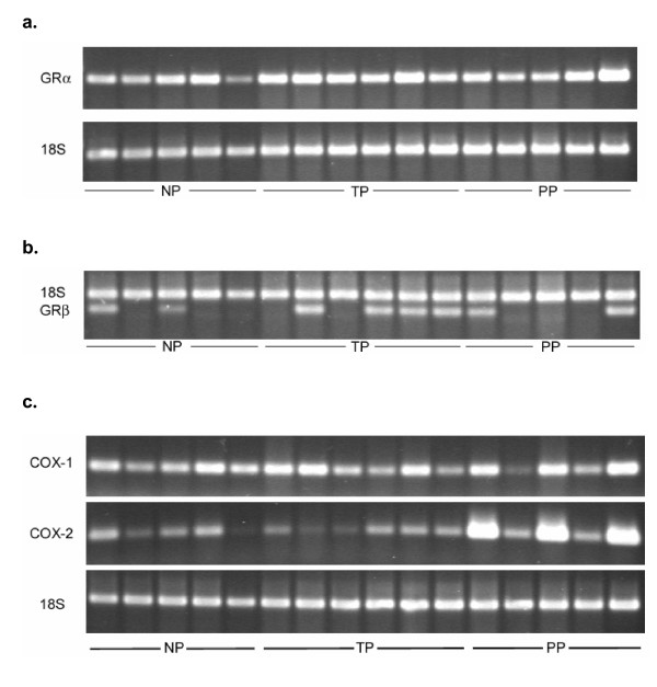
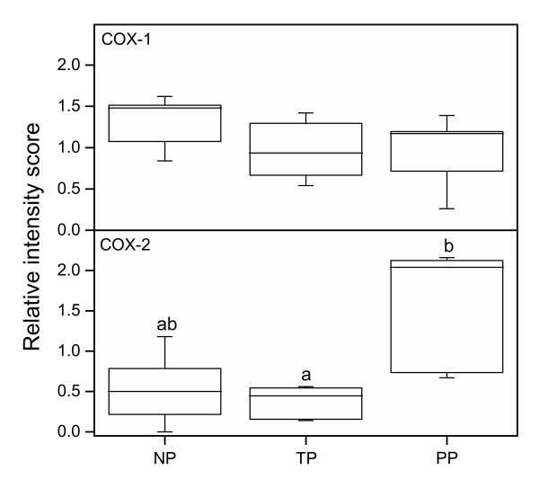
Methods: We monitored GR and NFkappaB, prostaglandin synthases cyclooxygenase (COX)-1 and -2, iNOS, as well as the PAF-receptor (PAF-R) in the uterine cervix from term pregnant women (with unripe cervices) before the onset of labor (TP), immediately after parturition (PP), as compared to non-pregnant (NP), using immunohistochemistry and RT-PCR.

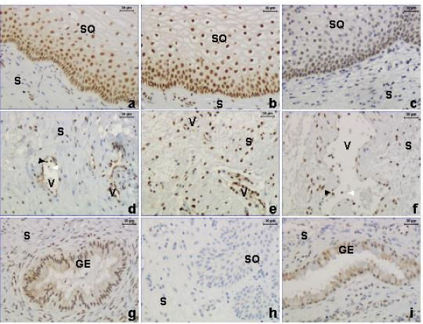
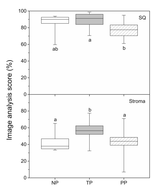
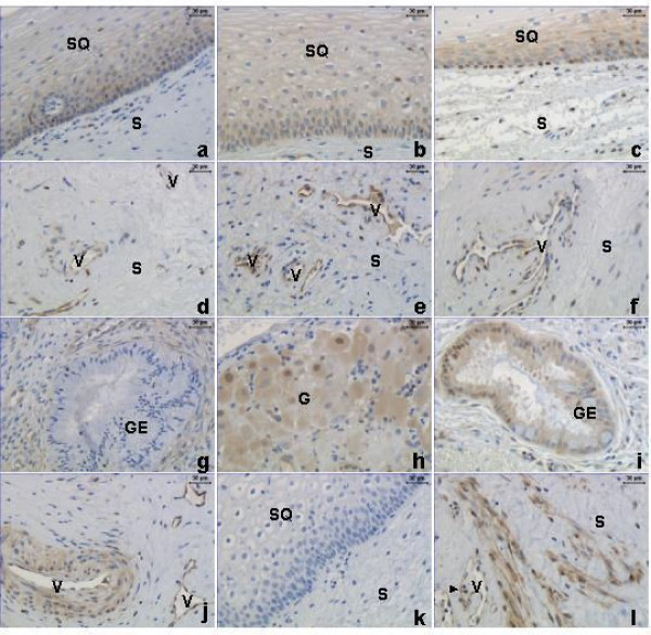
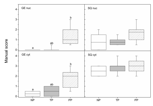
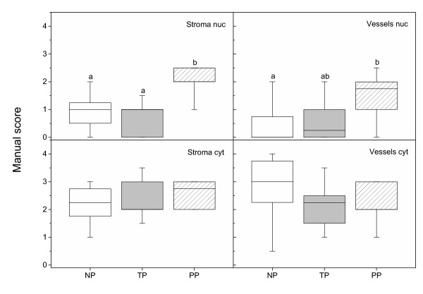
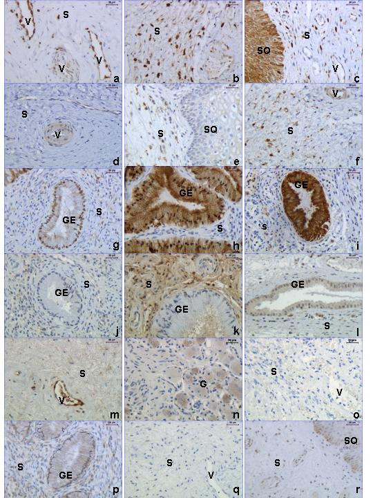
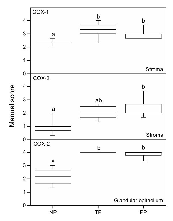
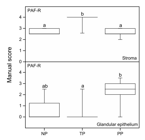
Results: The GR protein was detected by immunohistochemistry in the nuclei of stroma and squamous epithelium (SQ). Stromal GR staining was increased in TP as compared to the NP group and decreased again after parturition. GR staining in SQ was decreased after parturition as compared to term. NFkappaB was present in SQ and glandular epithelium (GE), stroma and vascular endothelium. Increased nuclear NFkappaB staining was observed postpartum as compared to term pregnancy in stroma and GE. Stromal immunostaining for COX-1 as well as COX-2 was increased in the TP and PP groups as compared to the NP, and GE displayed an intensely increased COX-2 immunostaining at term and postpartum. Stromal PAF-R immunostaining was highest at term, while it was greatly increased in GE postpartum. No difference in the immunostaining for iNOS was found between the groups. RT-PCR showed a predominance of GRalpha to GRbeta mRNA in cervical tissue. The COX-2 mRNA level was increased in the PP group as compared to the TP group.

Conclusions: There is a decrease in GR levels in human cervix at parturition. Concomitantly there is an increase of factors such as NFkappaB, PAF-R, COX-1 and COX-2, suggesting that they may participate in the sequence of events leading to the final cervical ripening.


Figures

Figure 1
Figure 1
Immunostaining of GR in the non-pregnant (NP) (left column, a, d, g), term pregnant (TP) (middle column, b, e, h) and postpartum (PP) (right column, c, f, i) groups. GR protein (brown staining) is present in the nuclei of cervical stroma (S), squamous epithelium (SQ), vessels (V) and glandular epithelium (GE) in all three groups. Some leukocytes, identified by their morphology, were positive for GR (black arrowheads) while others were negative (white arrowheads) (d and f). A negative control where the primary antibody was replaced by rabbit IgG is shown in h.
Figure 2
Figure 2
GR levels, as assessed by image analysis of immunohistochemistry results, in cervical squamous epithelium (SQ) and stroma in samples from TP (n = 12) and PP (n = 14) as compared to NP (n = 8) women. Box and whisker plots represent the median value with 50% of all data falling within the box. The "whiskers" extend to the 5th and 95th percentiles. Boxes with different letter designations are significantly different, p < 0.05.
Figure 3
Figure 3
Immunostaining of NFκB protein in stroma (S), glandular epithelium (GE), squamous epithelium (SQ) and vessels (V) in cervical biopsies from the NP (left column, a, d, g, j), TP (middle column, b, e, h) and PP (right column, c, f, i, l) groups. Positive nuclear and cytoplasmic immunostaining is also observed in neuronal ganglions (G) (h). Some leukocytes, identified by their morphology, display positive NFκB staining (l, black arrowhead). A negative control where the primary antibody is replaced by rabbit IgG is shown in k.
Figure 4
Figure 4
NFκB protein levels, as assessed by manual scoring of immunohistochemistry results, in nuclei and cytoplasma of glandular epithelium (GE) and squamous epithelium (SQ) in cervical samples from TP (n = 10) and PP (n = 10) as compared to NP (n = 8) women. Box and whisker plots represent the median value with 50% of all data falling within the box. The "whiskers" extend to the 5th and 95th percentiles. Boxes with different letter designations are significantly different, p < 0.05.
Figure 5
Figure 5
NFκB protein levels, as assessed by manual scoring, in nuclei and cytoplasma of stroma and vascular endothelium in cervical samples from TP (n = 10) and PP (n = 10) as compared to NP (n = 8) women. Box and whisker plots represent the median value with 50% of all data falling within the box. The "whiskers" extend to the 5th and 95th percentiles. Boxes with different letter designations are significantly different, p < 0.05.
Figure 6
Figure 6
Immunostaining of COX-1 (a-c), COX-2 (d-i), PAF-R (j-n) and iNOS (p, r), in cervices from women in the NP (left column), TP (middle column) and PP (right column) group. A representative negative control for COX-1, COX-2 and PAF-R immunohistochemistry (primary antibody replaced by goat IgG, same secondary antibody) is shown in o, the negative control for iNOS is shown in q (primary antibody replaced by mouse IgG). Abbreviations: S = stroma, GE = glandular epithelium, SQ = squamous epithelium, V = vessel and G = ganglion cells.
Figure 7
Figure 7
COX-1 (top) and COX-2 (middle, bottom) protein levels, as assessed by manual scoring of stroma (top, middle) and glandular epithelium (bottom) in cervical samples from the TP (n = 8), PP (n = 9) and NP (n = 6) groups. Box and whisker plots represent the median value with 50% of all data falling within the box. The "whiskers" extend to the 5th and 95th percentiles. Boxes with different letter designations are significantly different, p < 0.05.
Figure 8
Figure 8
PAF-R protein levels, as assessed by manual scoring, in stroma (top) and glandular epithelium (bottom) in cervical samples from the TP (n = 11), PP (n = 13) and NP (n = 9) groups. Box and whisker plots represent the median value with 50% of all data falling within the box. The "whiskers" extend to the 5th and 95th percentiles. Boxes with different letter designations are significantly different, p < 0.05.
Figure 9
Figure 9
Images of representative RT-PCR gels for a: GRα, b: GRβ and c: COX-1 (upper band) and COX-2 (middle band) in the human cervix in the non-pregnant (NP; n = 5), term pregnant (TP; n = 6) and postpartum (PP; n = 5) groups. The gels are stained with ethidium bromide. a: GRαmRNA(upper band) and 18S mRNA (lower band). b: 18S mRNA (upper band) and GRβmRNA(lower band). c: The 18S mRNA (bottom band) and the COX-1 (upper band) and COX-2 (middle band) PCR products.
Figure 10
Figure 10
COX-1 (top) and COX-2 (bottom) mRNA levels in the human cervix from the NP (n = 5), the TP (n = 6) and the PP (n = 5) groups as determined by RT-PCR. Intensities of the PCR product bands were normalized against the internal 18S standard. Box and whisker plots represent the median value with 50% of all data falling within the box. The whiskers extend to the 5th and 95th percentiles. Group medians with different letter superscripts are significantly different (P < 0.05).

References

    1. Ekman G, Malmström A, Uldbjerg N, Ulmsten U. Cervical collagen: an important regulator of cervical function in term labor. Obstet Gynecol. 1986;67:633–636. - PubMed
    1. Stygar D, Wang H, Stjernholm-Vladic Y, Ekman G, Eriksson H, Sahlin L. Co-localization of estrogen receptorβ and leukocyte markers in the human cervix. Mol Hum Reprod. 2001;7:881–886. doi: 10.1093/molehr/7.9.881. - DOI - PubMed
    1. Osman I, Young A, Ledingham MA, Thomson AJ, Jordan F, Greer IA, Norman JE. Leukocyte density and pro-inflammatory cytokine expression in human fetal membranes, decidua, cervix and myometrium before and during labour at term. Mol Hum Reprod. 2003;9:41–45. doi: 10.1093/molehr/gag001. - DOI - PubMed
    1. Sennström M, Ekman G, Westergren-Thorsson G, Malmström A, Byström B, Endresen U, Mlambo N, Norman M, Ståbi B, Brauner A. Human cervical ripening, an inflammatory process mediated by cytokines. Mol Hum Reprod. 2000;6:375–381. doi: 10.1093/molehr/6.4.375. - DOI - PubMed
    1. McKay LI, Cidlowski JA. Molecular control of immune/inflammatory responses: Interactions between nuclear factor-κB and steroid receptor signaling pathways. Endocrine Rev. 1999;20:435–459. doi: 10.1210/er.20.4.435. - DOI - PubMed

Publication types

MeSH terms