Comparative Study
. 2004 Nov 15;275(2):473-92.
doi: 10.1016/j.ydbio.2004.08.047.

Gene expression profiling of the developing Drosophila CNS midline cells

Affiliations
Comparative Study

Gene expression profiling of the developing Drosophila CNS midline cells

Joseph B Kearney et al. Dev Biol. .

Abstract

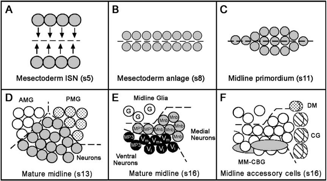
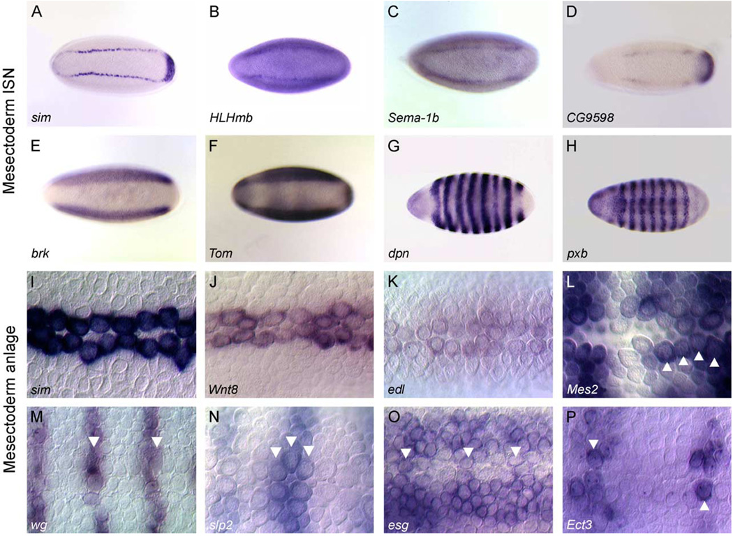
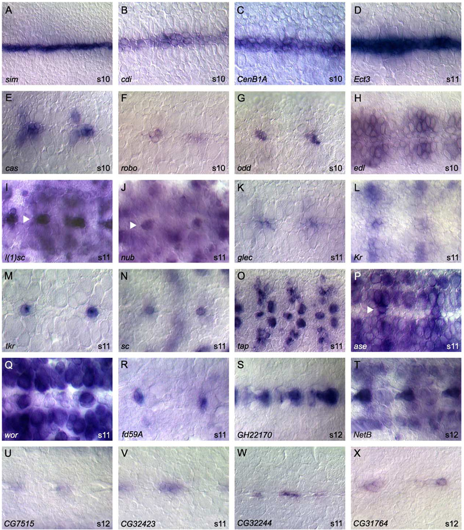
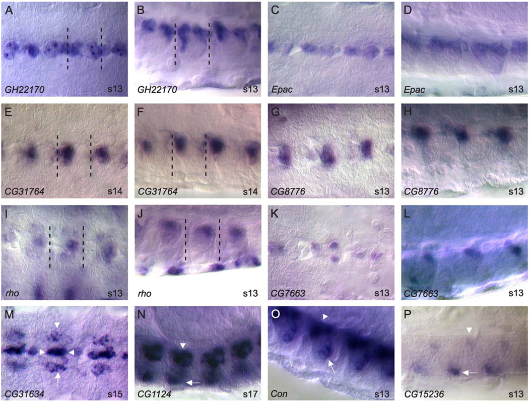
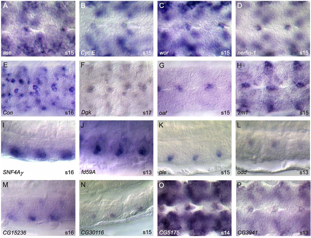
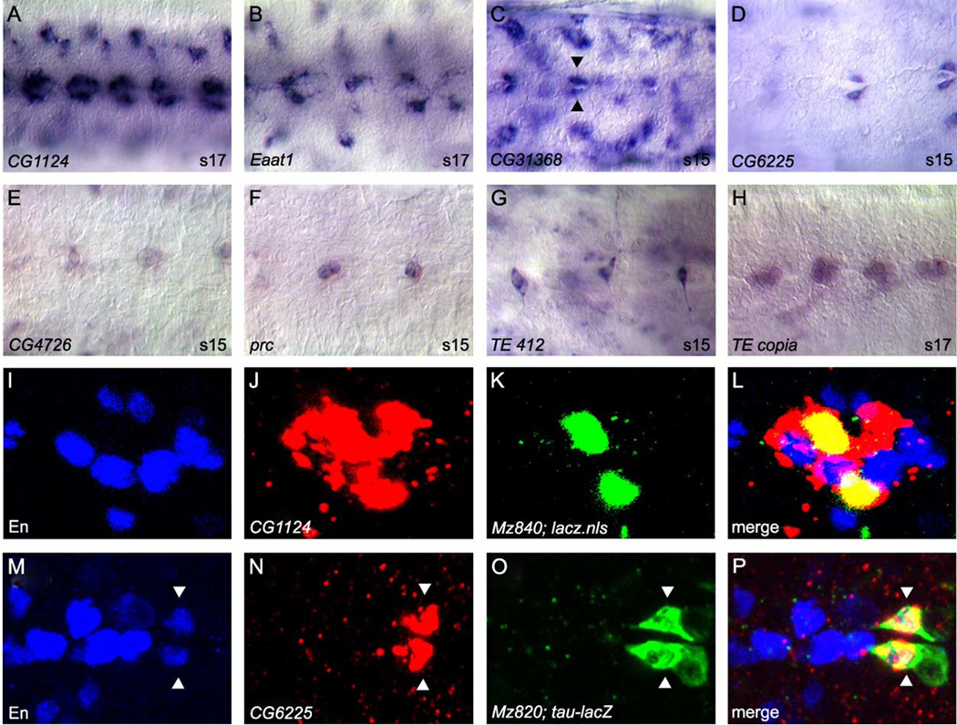
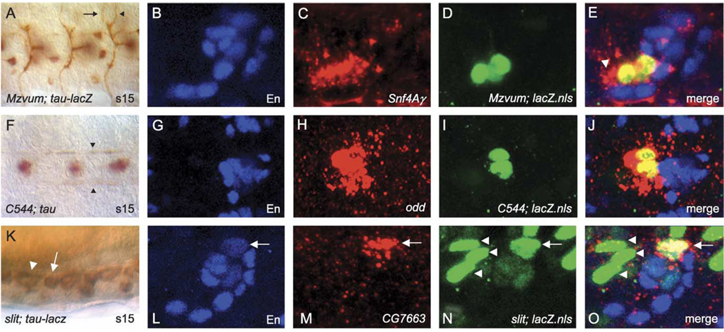
The Drosophila CNS midline cells constitute a specialized set of interneurons, motorneurons, and glia. The utility of the CNS midline cells as a neurogenomic system to study CNS development derives from the ability to easily identify CNS midline-expressed genes. For this study, we used a variety of sources to identify 281 putative midline-expressed genes, including enhancer trap lines, microarray data, published accounts, and the Berkeley Drosophila Genome Project (BDGP) gene expression data. For each gene, we analyzed expression at all stages of embryonic CNS development and categorized expression patterns with regard to specific midline cell types. Of the 281 candidates, we identified 224 midline-expressed genes, which include transcription factors, signaling proteins, and transposable elements. We find that 58 genes are expressed in mesectodermal precursor cells, 138 in midline primordium cells, and 143 in mature midline cells--50 in midline glia, 106 in midline neurons. Additionally, we identified 27 genes expressed in glial and mesodermal cells associated with the midline cells. This work provides the basis for future research that will generate a complete cellular and molecular map of CNS midline development, thus allowing for detailed genetic and molecular studies of neuronal and glial development and function.


Figures

Fig. 1
Fig. 1
Schematic summary of CNS midline cell development. In all panels, a single segment is shown with anterior to the left. Embryonic stages are indicated by “s#”. (A) Mesectoderm ISN stage, ventral view. Two stripes of mesectodermal cells reside on either side of the mesoderm in the blastoderm embryo (stage 5). Dotted line indicates ventral midline of embryo. There are four cells/segment on each side. Arrows represent how the mesectodermal cells move together at the ventral midline during gastrulation (stage 6) as the mesoderm invaginates. (B) Mesectoderm anlage stage, ventral view. During the mesectoderm anlage stage (stages 7–8), the mesectodermal cells meet at the midline and then undergo a synchronous cell division, resulting in 16 cells per segment. (C) Midline primordium stage, ventral view. During the midline primordium stage, midline cells rearrange from a two-cell wide planar array into a cell cluster. Midline cells within these clusters differ slightly in their dorsal/ventral positions. (D) Mature CNS midline cells, stage 13. Sagittal view, dorsal up. At stage 13, two populations of midline glial cells become evident. The anterior midline glia (AMG; open circles) are reduced by apoptosis but ultimately will ensheathe the commissures while all posterior midline glia (PMG; dotted circles) will undergo apoptosis. Midline neurons (shaded circles) occupy the space between and below the midline glia. Dotted lines separate the different cell groups. (E) Mature CNS midline cells, stage 16. Sagittal view, dorsal is up. The PMG have undergone apoptosis and are absent, whereas the AMG give rise to ~3 mature glia (G, open circles). Midline neurons have migrated to their final positions within the ganglion. Medial neurons include MP1 neurons (MP1, shaded circles) and the progeny of the MNB (Mnb, shaded circles). Ventral neurons include VUM motorneurons (Vm, black circles), VUM interneurons (Vi, black circles), and MP3 neurons (MP3, black circles). (F) Midline accessory cells shown in relation to midline neurons and glia (open circles). Two DM cells (dotted circles) lie atop the CNS near the midline channel, which is lined by six-channel glia (CG; hatched ovals). The two MM-CBG in each segment (shaded ovals) are closely associated with the ventral neurons.
Fig. 2
Fig. 2
Gene expression during the mesectoderm ISN and mesectoderm anlage stages. Whole-mount embryos hybridized in situ showing the expression patterns of genes expressed in: (A–H) mesectoderm ISN, and (I–P) mesectoderm anlage. All views are ventral with anterior to the left. Gene names are shown at the bottom left. Representative genes are shown for three classes of mesectoderm ISN expression: (A–D) genes with relatively specific expression in all mesectoderm ISN cells, (E–F) genes expressed in all mesectoderm ISN cells and showing additional expression in other neuroectodermal cells, and (G–H) genes with pair-rule patterns expressed in subsets of mesectoderm ISN cells. (I–P) Shown are genes expressed in: (I–K) all mesectodermal anlage cells, and (L–P) subsets of mesectodermal anlage cells. Arrowheads indicate stained mesectodermal cells. Note the number of mesectoderm anlage cells labeled per hemisegment varies for different genes. Magnification is 12.5× for A–H and 80× for I–P.
Fig. 3
Fig. 3
Midline primordium gene expression. Whole-mount stages 9–12 embryos hybridized to RNA probes. All views are ventral with anterior to the left. Arrowheads denote midline cells in embryos with high surrounding expression. Representatives of two major classes of gene expression are shown: (A–D) expression in all midline cells, and (E–X) expression in subsets of midline cells. Genes expressed in subsets can be further divided into those expressed in: (E–L) multiple primordium cells, and (M–P) single primordium cells. (Q–T) A subset of genes expressed in the primordium stages maintains expression in (Q–R) mature midline neurons or (S–T) midline glia. Compare (S) GH22170 in the primordium to GH22170 in the mature midline glia (Figs. 4A–B), and (Q) wor in the primordium to wor in midline neurons (Fig. 5C). (U–X) Shown are genes of currently uncharacterized function expressed in subsets of midline primordium cells. Magnification is 80× for all except Q (100×).
Fig. 4
Fig. 4
Midline glial gene expression. Whole-mount stages 13–17 embryos hybridized to RNA probes. (A–L) Each gene is shown twice with a ventral view on the left and sagittal view on the right. Anterior is left and dorsal top for sagittal views. Dotted lines enclose a single ganglion. (A-D) Two genes expressed in both anterior and posterior midline glia. (E–H) Two genes expressed prominently in anterior midline glia. (I–L) Two genes expressed prominently in posterior midline glia. (M–P) Ventral (M) or sagittal (N–P) views of embryos hybridized with probes to genes expressed in midline glia and other cell types. (M) CG31634 is expressed in both midline glia (arrowhead) and longitudinal glia (arrow). (N) CG1 124 is expressed in both midline glia (arrowhead) and MM-CBG (arrow). (O–P) Examples of genes expressed in both midline glia (arrowhead) and midline neurons (arrow). Magnification is 80x.
Fig. 5
Fig. 5
Midline neuronal expression. Whole-mount stages 13–17 embryos hybridized to RNA probes. Anterior is to the left and dorsal top for sagittal views. (A–H) Ventral views of genes expressed in: (A–D) medial neurons, and (E–H) ventral neurons. (I–L) Sagittal views showing the dorsal/ventral position of genes expressed in (I–K) ventral neurons and (L) medial neurons. (M–P) Examples genes of uncharacterized function expressed in (M–N) ventral neurons (sagittal views), and (O–P) medial neurons (ventral views). Magnification is 80x.
Fig. 6
Fig. 6
Midline accessory cell gene expression and midline-expressed transposable elements. (A–H) Whole-mount stage 15 or 17 embryos were hybridized to RNA probes and visualized by AP/DIC imaging. All views are ventral with anterior to the left. Magnification is 80x. (A–G) Examples of genes expressed in subsets of midline accessory cells, including (A–B) MM-CBG, (C–D) channel glia (arrowheads), and (E–G) DM cells. (G–H) Transposable element expression in midline and accessory cells, including (G) DM cells and (H) ventral neurons. (I–P) Confocal images of multi-label fluorescent in situ hybridization. An individual ganglion is shown for each panel. All views are ventral with anterior to the left. (I–L) Stage 16 Mz840-Gal4 × UAS-GFP-lacZ.nls embryo co-labeled with: (I) anti-En, (J) CG1124 RNA probe, and (K) anti-β-gal. (L, merge image) CG1124 expression (red) colocalizes with Mz840- driven nuclear β-gal (green) expression in MM-CBG. MM-CBGs are positioned just lateral to En-positive VUM cells. The in situ fluorescence stains the cytoplasm and is usually broader than the nuclear staining observed with immunostaining of GFP-lacZ.nls or nuclear proteins. (M–P) Stage 15 Mz820-Gal4 × UAS-tau-lacZ embryo co-labeled with: (M) anti-En, (N) CG6225 RNA probe, and (O) anti-β-gal. (P, merge image) CG6225 expression (red) co-localizes with tau-lacZ fusion protein (green) expressed in channel glia (arrowheads) via the Mz820-Gal4 enhancer trap line. CG6225-positive channel glia also express en.
Fig. 7
Fig. 7
Cellular correspondence of midline gene expression. (A, F, K) DIC images of Gal4×UAS-tau or UAS-tau-lacZ embryos immunostained with anti-Tau or anti-β-gal. (B–E, G–J, L–O) Confocal images showing midline cell co-localization of RNA probes with defined midline cell markers. Anterior is to the left in all cases, and for sagittal views, dorsal is up. (A) Ventral view of stage 15 MzVUM-Gal4×UAS-tau-lacZ embryo showing the VUM motorneuron axonal trajectories that project out the intersegmental (arrow) and segmental (arrowhead) nerve roots. The VUM motorneuron cell bodies are out of the plane of focus. (B–E) Sagittal view of an individual ganglion from a MzVUM-Gal4×UAS-GFP-lacZ.nls embryo stained for (B) anti-En (blue), (C) Snf4Ay RNA (red), (D) anti-β-gal (green), and (E) merge image showing that Snf4Ay RNA co-localizes with β-gal-stained VUM motorneurons. Additional expression is detected just anterior to VUM motorneurons (arrowhead) and may represent staining in MP3 neurons. (F) Ventral view of a stage 15 C544-Gal4×UAS-tau embryo stained with anti-Tau showing the MP1 axonal trajectories along the longitudinal connectives (arrowheads). (G–J) Ventral view of an individual ganglion from a stage 16 C544-Gal4×UAS-GFP-lacZ.nls embryo stained for (G) anti-En (blue), (H) odd RNA (red), (I) anti-β-gal (green), and (J) merge image showing that odd RNA co-localizes with β-gal-stained MP1 neurons. (K) Sagittal view of stage 15 slit-gal4×UAS-tau-lacZ embryo showing anterior (arrowhead) and posterior (arrow) glia within a single ganglion. (L–O) Sagittal view of a single ganglion from a slit-Gal4×UAS-GFP-lacZ.nls embryo stained for (L) anti-En (blue), (M) CG7663 RNA (red), (N) anti-β-gal (green), and (O) merge image showing that CG7663 RNA co-localizes with β-gal and En-stained posterior glia (arrow). Posterior glia (arrow) can be easily distinguished from the three anterior glia (arrowheads) based on their relative positions along the A–P axis.

Similar articles

Cited by

References

    1. Araujo SJ, Tear G. Axon guidance mechanisms and molecules: lessons from invertebrates. Nat. Rev. Neurosci. 2003;4:910–922. - PubMed
    1. Ashraf SI, Ip YT. The Snail protein family regulates neuroblast expression of inscuteable and string, genes involved in asymmetry and cell division in Drosophila. Development. 2001;128:4757–4767. - PubMed
    1. Ashraf SI, Hu X, Roote J, Ip YT. The mesoderm determinant snail collaborates with related zinc-finger proteins to control Drosophila neurogenesis. EMBO J. 1999;18:6426–6438. - PMC - PubMed
    1. Bate CM, Grunewald EB. Embryogenesis of an insect nervous system II: a second class of neuron precursor cells and the origin of the intersegmental connectives. J. Embryol. Exp. Morphol. 1981;61:317–330. - PubMed
    1. Bennetzen JL. Transposable element contributions to plant gene and genome evolution. Plant Mol. Biol. 2000;42:251–269. - PubMed

Publication types

MeSH terms

LinkOut - more resources