. 2004 Nov;72(11):6211-20.
doi: 10.1128/IAI.72.11.6211-6220.2004.

Passive immunization of neonatal mice against Pneumocystis carinii f. sp. muris enhances control of infection without stimulating inflammation

Affiliations

Passive immunization of neonatal mice against Pneumocystis carinii f. sp. muris enhances control of infection without stimulating inflammation

Kerry M Empey et al. Infect Immun. 2004 Nov.

Abstract

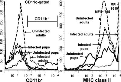
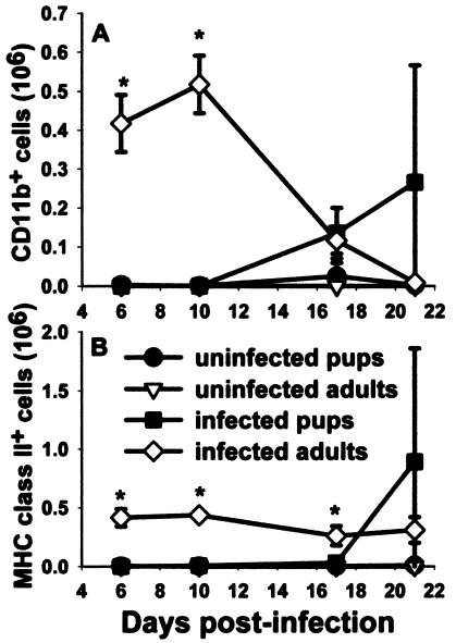
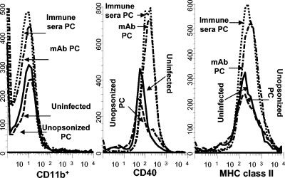
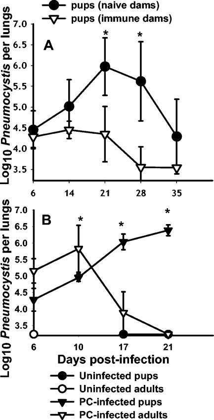
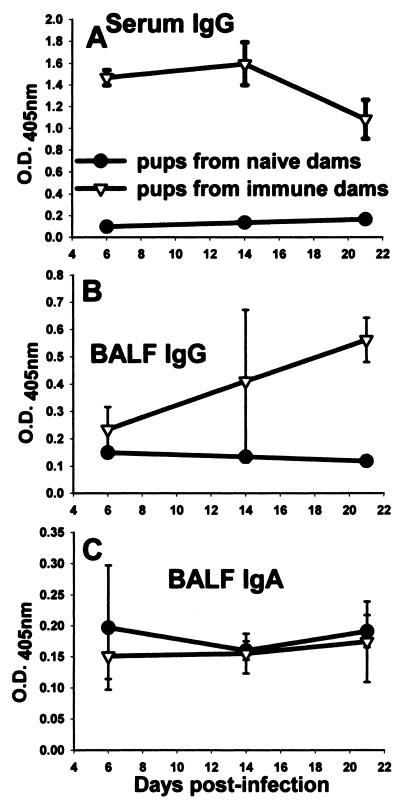
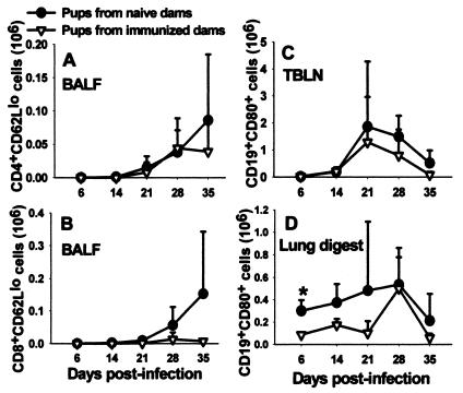
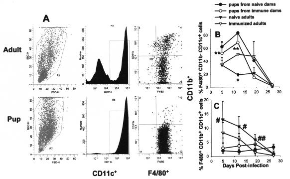
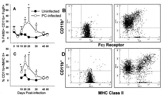
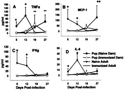
Pneumocystis carinii is an opportunistic fungal pathogen that causes life-threatening pneumonia in immunocompromised individuals. Infants appear to be particularly susceptible to infection with Pneumocystis. We have previously shown that there is a significant delay in clearance of the organisms from the lungs of neonatal mice compared to adults. Since alveolar macrophages are the effector cells responsible for killing and clearance of Pneumocystis, we have examined alveolar macrophage activity in neonatal mice. We found that alveolar macrophage activation is delayed about 1 week in Pneumocystis-infected neonates compared to adults. Opsonization of the organism by Pneumocystis-specific antibody resulted in increased clearance of the organism in neonatal mice; however, there was decreased expression of activation markers on neonatal alveolar macrophages and reduced levels of cytokines associated with macrophage activation. Mice born to immunized dams had significant amounts of Pneumocystis-specific immunoglobulin G in their lungs and serum at day 7 postinfection, whereas mice born to naive dams had merely detectable levels. This difference correlated with enhanced Pneumocystis clearance in mice born to immunized dams. The increase in specific antibody, however, did not result in significant inflammation in the lungs, as no differences in numbers of activated CD4+ cells were observed. Furthermore, there was no difference in cytokine or chemokine concentrations in the lungs of pups born to immune compared to naive dams. These findings indicate that specific antibody plays an important role in Pneumocystis clearance from lungs of infected neonates; moreover, this process occurs without inducing inflammation in the lungs.


Figures

FIG. 1.
FIG. 1.
Expression levels of large nonlymphocytes of CD11b and MHC class II from alveolar spaces of Pneumocystis-infected animals were very low at day 10 postinfection in neonatal mice compared to those for adult mice. Mice were infected with Pneumocystis as neonates (24 to 72 h after birth) or adults, and lungs were lavaged at day 10 postinfection to examine macrophage levels. Cells were stained with antibodies specific for CD11c and CD11b or MHC class II, and phenotypes were examined by using flow cytometry. Representative histograms of CD11b-positive cells (gated on CD11c+ cells) and MHC class II-positive cells (gated on large nonlymphocytes) are shown. Cells were gated for large nonlymphoid cells by using forward and side light scatter. Controls included BALF cells from uninfected neonates or adults. Data are representative of results for three to five mice per group.
FIG. 2.
FIG. 2.
Numbers of nonlymphoid cells expressing CD11b and MHC class II in the alveolar spaces of Pneumocystis-infected neonates were significantly lower than those in adult lungs through 2 weeks postinfection. Mice were infected with Pneumocystis as neonates (24 to 72 h after birth) or adults. Control mice received HBSS vehicle. Lungs were lavaged at the indicated time points, cells were stained with antibodies specific for CD11b and MHC class II, and cells were examined by using flow cytometry. Cells were gated on large nonlymphocytes, and the numbers of cells expressing (A) CD11b and (B) MHC class II were counted. Data represent the means ± SD for three to five mice per group. *, P < 0.05 compared to all other groups at the same time point.
FIG. 3.
FIG. 3.
No difference was observed in the expression of MHC class II, CD40, and CD11b on alveolar lymphocytes isolated 10 days postinfection in mice treated with opsonized Pneumocystis (PC). Neonatal mice were infected with unopsonized Pneumocystis or organisms were pretreated with polyclonal antiserum or MAb 4F11(G1). A control group was uninfected. Lung lavage cells were stained with antibodies specific for MHC class II, CD40, or CD11b, and the phenotypes of large nonlymphoid cells were determined by flow cytometry. Data are histograms of nonlymphoid gated cells from individual mice at day 10 postinfection and are representative of results from three to four mice per group.
FIG. 4.
FIG. 4.
Control of Pneumocystis infection in neonates is expedited in the presence of maternal antibody; however, it still lags behind clearance in adult mice. (A) Adult female mice were challenged with i.t. inoculations of Pneumocystis (PC) on the day they were placed into cages with males for breeding. They were given a boost at day 16 of gestation. Control dams were inoculated with PBS. Neonatal mice were infected with Pneumocystis within 72 h of birth, and lung burden was determined at the indicated time points. Data are expressed as the means ± SD for four to five mice per group. *, P < 0.05 compared to pups from immunized dams. (B) Adult and neonatal mice were given i.n. inoculations of Pneumocystis (PC), and the numbers of organisms were detected microscopically over time. Control mice were given inoculations of PBS. Data represent the means ± SD for four to five mice per group. *, P < 0.05 compared to infected pups.
FIG. 5.
FIG. 5.
Pups born to immunized dams had significant levels of Pneumocystis-specific IgG in the serum and BALF. Dams were given i.t. inoculations of Pneumocystis on the day of breeding and boosted with i.n. inoculums on day 16 of gestation. Control dams received inoculations of PBS. Pups in both groups were given i.n. inoculations of Pneumocystis within 72 h of birth. ELISAs were performed on BALF and serum collected from pups in each group to determine the level Pneumocystis-specific (A, B) IgG or (C) IgA present. Data are expressed as the optical density at 405 nm. BALF samples were undiluted and serum samples were diluted 1:100 for the ELISA assay. Data represent the means ± SD for four to five mice per group. *, P < 0.05 compared to pups from naïve dams.
FIG. 6.
FIG. 6.
No differences in the number of activated CD4+ and CD8+ or B cells in the lungs or draining lymph nodes were observed between pups born to either immunized or naïve dams. Dams were given i.t. inoculations of Pneumocystis on the day of breeding and boosted with i.n. inoculums on day 16 of gestation. Control dams received inoculations of PBS. Pups in both groups were then given i.n. inoculations of Pneumocystis within 72 h of birth. Activated T cells in the BALF (A, B) and B cells in the TBLN (C) and digested lung tissue (D) were determined by using fluorochrome-labeled antibodies specific for CD4, CD8, CD62L, CD19, and CD80 and flow cytometry at days 6, 14, 21, 28, and 35. Data represent the means ± SD for three to five mice per group.
FIG. 7.
FIG. 7.
The phenotype of alveolar macrophages was not changed in pups born to immune dams versus naïve dams over time. Dams were given i.t. inoculations of Pneumocystis on the day of breeding and boosted with i.n. inoculums on day 16 of gestation. Control dams received inoculations of PBS. Pups in both groups were then given i.n. inoculations of Pneumocystis within 72 h of birth. BALF was collected at the indicated time points postinfection, and cells were stained for macrophage and activation markers including F4/80, CD11b, CD11c, and MHC class II and analyzed by flow cytometry. (A) BALF flow cytometry data from an individual Pneumocystis-infected naïve adult and a pup from a naïve dam at day 12 postinfection. Forward and side scatters are shown in the left panels. The center panels show CD11c histograms of cells in the large cell region of the scatter profiles. The dot plots on the right side are F4/80 and CD11b staining of the CD11c-positive cells. Proportions of F4/80+ CD11b CD11c+ (B) and F4/80+ CD11b+ CD11c+ (C) alveolar macrophages present in the BALF of Pneumocystis-infected adult mice compared to neonates are shown. Data represent the means ± SD for four to five mice per group. *, P < 0.05 compared to all other groups at the same time point; **, P < 0.05 compared to adult groups at the same time point; #, P < 0.05 compared to pup groups at the same time point; ##, P < 0.05 compared to immunized adults at the same time point.
FIG. 8.
FIG. 8.
The pattern of expression of FcγR changed in mice infected with Pneumocystis as neonates. Neonatal mice (48 to 72 h old) were given i.n. inoculations of Pneumocystis or given a sham inoculation of buffer. BALF was collected at the indicated time points postinfection, and cells were stained for macrophage and activation markers including F4/80, CD11b, CD11c, and either FcγR or MHC class II and analyzed by flow cytometry. The proportions of F4/80+ CD11b+ FcγR+ cells (A) and F4/80+ CD11b+ MHC class II-positive cells (C) in BALF over time postinfection are shown. Dot plots showing CD11b versus either FcγR (B) or MHC class II (D) expression in individual mice at day 21 postinfection are shown. Data were gated on nonlymphocytes and F4/80+ cells. Panels on the left show data from a representative uninfected mouse, and panels on the right show data from a Pneumocystis-infected mouse. Data represent the means ± SD for four to five mice per group. *, P < 0.05 compared to pups at the same time point.
FIG. 9.
FIG. 9.
No differences in the concentrations of inflammatory cytokines were seen in BALF of Pneumocystis-infected pups born to naïve dams compared to those seen in pups born to immune dams through day 19 postinfection. Dams were given i.t. inoculations of Pneumocystis on the day of breeding and boosted with i.n. inoculums on day 16 of gestation. Control dams received inoculations of PBS. Pups in both groups were then given i.n. inoculations of P. carinii within 72 h of birth. BALF was collected from pups from immunized and naïve dams and from immunized and naïve adult mice. TNF-α, IFNγ, MCP-1, and IL-6 (panels A, B, C, and D, respectively) concentrations were determined by flow cytometry using cytometric bead arrays. Data represent the means ± SD for four to five mice per group. *, P < 0.05 compared to all other groups at the same time point, **, P < 0.05 compared to adult groups at the same time point.

Similar articles

Cited by

References

    1. Adkins, B. 1999. T-cell function in newborn mice and humans. Immunol. Today 20:330-335. - PubMed
    1. Adkins, B., and R.-Q. Du. 1998. Newborn mice develop balanced Th1/Th2 primary effector responses in vivo but are biased to Th2 secondary responses. J. Immunol. 160:4217-4224. - PubMed
    1. Adkins, B., and K. Hamilton. 1992. Freshly isolated, murine neonatal T cells produce IL-4 in response to anti-CD3 stimulation. J. Immunol. 149:3448-3455. - PubMed
    1. Bartlett, M. S., S. H. Vermund, R. Jacobs, P. J. Durant, M. M. Shaw, J. W. Smith, X. Tang, J. J. Lu, B. Li, S. Jin, and C.-J. Lee. 1997. Detection of Pneumocystis carinii DNA in air samples: likely environmental risk to susceptible persons. J. Clin. Microbiol. 35:2511-2513. - PMC - PubMed
    1. Berclaz, P. Y., Y. Shibata, J. A. Whitsett, and B. C. Trapnell. 2002. GM-CSF, via PU.1, regulates alveolar macrophage FcγR-mediated phagocytosis and the IL-18/IFN-γ-mediated molecular connection between innate and adaptive immunity in the lung. Blood 100:4193-4200. - PubMed

Publication types

MeSH terms