. 2004 Nov;72(11):6255-61.
doi: 10.1128/IAI.72.11.6255-6261.2004.

Recognition of mycobacterial antigens delivered by genetically detoxified Bordetella pertussis adenylate cyclase by T cells from cattle with bovine tuberculosis

Affiliations

Recognition of mycobacterial antigens delivered by genetically detoxified Bordetella pertussis adenylate cyclase by T cells from cattle with bovine tuberculosis

H Martin Vordermeier et al. Infect Immun. 2004 Nov.

Abstract

The exponential increase in the incidence of tuberculosis in cattle over the last two decades in the British national herd constitutes a significant economic problem. Therefore, research efforts are under way to develop cattle tuberculosis vaccines and specific diagnostic reagents to allow the distinction of vaccinated from infected animals. Mycobacterial antigens like ESAT-6 and CFP10 allow this distinction. This study investigates whether fusion protein of ESAT-6 or CFP10 with genetically detoxified Bordetella pertussis adenylate cyclase (CyaA) are recognized by Mycobacterium bovis-infected cattle more effectively than conventional recombinant proteins are, thus enhancing sensitivity or reducing the amount of antigens required. By measuring the frequencies of gamma interferon (IFN-gamma)-producing cells, we were able to show that the presentation of CFP10 as a CyaA fusion protein enhanced the molecular efficiency of its recognition 20-fold, while the recognition of ESAT-6 was not improved by CyaA delivery. Furthermore, in the whole-blood IFN-gamma test currently used in the field, the delivery of CFP10 and ESAT-6 by fusion to CyaA increased the amount of IFN-gamma produced and hence the proportion of infected animals responding to CFP10. The improved T-cell recognition of CyaA336/CFP10 was found to be dependent upon interaction with CD11b. In addition, presentation of CyaA336/CFP10 to CD4+ T cells was chloroquine sensitive, and CFP10 delivery by CyaA resulted in its accelerated presentation to T cells. In conclusion, the use of CyaA fusion proteins with ESAT-6 and CFP10 has the potential to improve the sensitivity of immunodiagnostic tests detecting bovine tuberculosis in cattle.


Figures

FIG. 1.
FIG. 1.
Dose-response relationship of in vitro IFN-γ production after stimulation with CFP10 (squares) and CyaA336/CFP10 (triangles). The readout system was the IFN-γ ELISPOT assay. SFC numbers from cultures with medium alone were subtracted from all values. In addition, the numbers of spots after incubation with CyaA alone were subtracted from the SFC induced by CyaA336/CFP10 stimulation. Tests were performed in duplicate with 2 × 10⁵ PBMC/well isolated from an M. bovis-infected calf. Horizontal lines indicate the maximum SFC numbers (peak values) induced after stimulation with CyaA336/CFP10 (a) and CFP10 (b); line c indicates the half-maximum SFC induced after CFP10 stimulation (50% maximum values). Vertical lines indicate the CyaA336/CFP10 (d) and CFP10 (e) concentrations required to induce 50% of CFP10-induced peak responses (50% maximum concentration).
FIG. 2.
FIG. 2.
Comparison of abilities of CyaA fusion proteins and recombinant proteins to stimulate in vitro IFN-γ production by PBMC from M. bovis-infected cattle. Left panel: 50% maximum concentrations determined as illustrated in Fig. 1. Right panel: peak values determined as illustrated in Fig. 1. The readout system was the IFN-γ ELISPOT assay. SFC numbers from cultures with medium alone were subtracted from all values. In addition, the numbers of spots after incubation with CyaA alone were subtracted from the SFC induced by CyaA336/CFP10 or CyaA336/ESAT-6 stimulation. Tests were performed in duplicate with 2 × 10⁵ PBMC/well. *, P < 0.05 (two-tailed Wilcoxon signed rank matched pairs test).
FIG. 3.
FIG. 3.
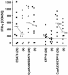
Performance of CyaA fusion proteins and recombinant ESAT-6 and CFP10 in the whole-blood Bovigam IFN-γ assay. Heparinized blood from eight M. bovis-infected calves was incubated with the antigens at 4 and 20 nM test concentrations. IFN-γ in plasma culture supernatants was determined by ELISA. The results are expressed as ΔOD450 units (OD450 × 1,000) with ΔOD450 values from cultures with medium alone subtracted from all values. In addition, the ΔOD450 values after incubation with CyaA alone were subtracted from wells with CyaA336/CFP10 or CyaA336/ESAT-6 stimulation. The horizontal line indicates cutoff for positivity (Δ125 OD450 units). Cultures were performed in duplicate in 96-well flat-bottomed plates. *, P < 0.05; **, P < 0.01 (two-tailed Wilcoxon signed rank matched pairs test).
FIG. 4.
FIG. 4.
Involvement of CD11b in the recognition of CyaA336/CFP10. Cultures were performed in the presence of a blocking CD11b-specific IgG1 MAb (ILA15) and an isotype control (CC94). The readout system was the IFN-γ ELISPOT assay. SFC numbers from cultures with medium alone were subtracted from all values. Tests were performed in duplicate with 2 × 10⁵ PBMC/well isolated from one infected calf. Responses are significantly different (P < 0.02) for each concentration tested except lowest concentration (one-tailed Wilcoxon signed rank matched pairs test).
FIG. 5.
FIG. 5.
Accelerated presentation kinetics of CyaA336/CFP10 to CD4+ T cells. CyaA336/CFP10 and CFP10 (both at 3.7 nM) were incubated with CD4-depleted PBMC as a source of APC. At the indicated time points, chloroquine was added to stop further processing. APC were then cultured for 48 h in the presence of 2 × 10⁴ cells from a CFP10-specific CD4+-T-cell line added. IFN-γ in culture supernatants was determined by ELISA. Triangles, CFP10; circles, CyaA336/CFP10; open symbols, no addition of chloroquine; closed symbols, chloroquine was added at indicated time points. Box, addition of chloroquine and antigens simultaneously. Results are expressed as means of triplicate determinations ± standard errors.

Similar articles

Cited by

References

    1. Accapezzato, D., R. Nisini, M. Paroli, G. Bruno, F. Bonino, M. Houghton, and V. Barnaba. 1998. Generation of an MHC class II-restricted T cell epitope by extracellular processing of hepatitis delta antigen. J. Immunol. 160:5262-5266. - PubMed
    1. Behr, M. A., M. A. Wilson, W. P. Gill, H. Salamon, G. K. Schoolnik, S. Rane, and P. M. Small. 1999. Comparative genomics of BCG vaccines by whole-genome DNA microarray. Science 284:1520-1523. - PubMed
    1. Buddle, B., N. A. Parlane, D. L. Keen, F. E. Aldwell, J. M. Pollock, K. Lightbody, and P. Andersen. 1999. Differentiation between Mycobacterium bovis BCG-vaccinated and M. bovis-infected cattle by using recombinant mycobacterial antigens. Clin. Diagn. Lab. Immunol. 6:1-5. - PMC - PubMed
    1. Buddle, B. M., A. R. McCarthy, T. J. Ryan, J. M. Pollock, H. M. Vordermeier, R. G. Hewinson, P. Andersen, and G. W. de Lisle. 2003. Use of mycobacterial peptides and recombinant proteins for the diagnosis of bovine tuberculosis in skin test-positive cattle. Vet. Rec. 153:615-620. - PubMed
    1. Cosivi, O., J. M. Grange, C. J. Daborn, M. C. Raviglione, T. Fujikura, D. Cousins, R. A. Robinson, H. F. Huchzermeyer, I. de Kantor, and F. X. Meslin. 1998. Zoonotic tuberculosis due to Mycobacterium bovis in developing countries. Emerg. Infect. Dis. 4:59-70. - PMC - PubMed

Publication types

MeSH terms