. 2004 Nov;72(11):6313-7.
doi: 10.1128/IAI.72.11.6313-6317.2004.

Functional analysis of Bacillus anthracis protective antigen by using neutralizing monoclonal antibodies

Affiliations

Functional analysis of Bacillus anthracis protective antigen by using neutralizing monoclonal antibodies

Fabien Brossier et al. Infect Immun. 2004 Nov.

Abstract

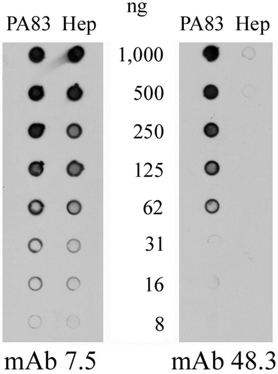
Protective antigen (PA) is central to the action of the lethal and edema toxins produced by Bacillus anthracis. It is the common cell-binding component, mediating the translocation of the enzymatic moieties (lethal factor [LF] and edema factor) into the cytoplasm of the host cell. Monoclonal antibodies (MAbs) against PA, able to neutralize the activities of the toxins in vitro and in vivo, were screened. Two such MAbs, named 7.5 and 48.3, were purified and further characterized. MAb 7.5 binds to domain 4 of PA and prevents the binding of PA to its cell receptor. MAb 48.3 binds to domain 2 and blocks the cleavage of PA into PA63, a step necessary for the subsequent interaction with the enzymatic moieties. The epitope recognized by this antibody is in a region involved in the oligomerization of PA63; thus, MAb 48.3 does not recognize the oligomer form. MAbs 7.5 and 48.3 neutralize the activities of anthrax toxins produced by B. anthracis in mice. Also, there is an additive effect between the two MAbs against PA and a MAb against LF, in protecting mice against a lethal challenge by the Sterne strain. This work contributes to the functional analysis of PA and offers immunotherapeutic perspectives for the treatment of anthrax disease.


Figures

FIG. 1.
FIG. 1.
Lethal toxin-neutralizing activity of anti-PA MAbs 7.5 and 48.3 on RAW264.7 macrophages. MAbs 7.5 (•) and 48.3 (▪) were preincubated with PA83, and the complexes were further incubated in the presence of LF with RAW264.7 cells. The viability of the cells was then assessed by a colorimetric assay. Results are expressed as the percentages of cells still viable. The experiment was carried out at least three times for each MAb with less than 10% of variability.
FIG. 2.
FIG. 2.
PA83 domains recognized by the MAbs. (A) Schematic representation of fragments generated by cleavage of PA83 by chymotrypsin. Domain 1 (residues 1 to 258), domain 2 (residues 259 to 487), domain 3 (residues 488 to 595), and domain 4 (residues 596 to 735) are shown. (B) Chymotrypsin digestion fragments of PA83 (each, 1 μg) were subjected to electrophoresis and were transferred to a nitrocellulose membrane for Western blotting with MAbs 7.5 and 48.3 as first antibodies. (C) PA83 and PA83 proteins with mutations in domain 4 (PA608, PA705, and PA711) were absorbed onto a nitrocellulose membrane for a dot blot experiment with MAbs 7.5 and 48.3 as first antibodies.
FIG. 3.
FIG. 3.
Inhibition of the initial steps of the intoxication process. MAbs 7.5 and 48.3 were separately incubated with radiolabeled PA83 and then added to CHO-K1 cells at 4°C. Membrane proteins were solubilized and subjected to electrophoresis. The presence of PA83 at the surface of the cells was revealed by autoradiography. The experiment was carried out at least three times for each MAb.
FIG. 4.
FIG. 4.
Purified PA83 or heptamer (Hep) of PA63 were diluted (from 1,000 to 8 ng) and adsorbed onto a nitrocellulose membrane for dot blotting with MAbs 7.5 and 48.3 as first antibodies. The experiment was carried out at least three times for each MAb.
FIG. 5.
FIG. 5.
Protection of mice conferred by MAbs against challenge with Sterne strain spores. MAbs, alone or in combination as indicated, were administered intravenously to mice at doses per animal of 100 μg (white bars), 10 μg (hatched bars), and 1 μg (black bars). Results are expressed as the percentage of animals surviving challenge with the Sterne strain. Each point is the mean of at least two duplicate experiments.

Similar articles

Cited by

References

    1. Abrami, L., S. H. Liu, P. Cosson, S. H. Leppla, and F. G. van der Goot. 2003. Anthrax toxin triggers endocytosis of its receptor via a lipid raft-mediated clathrin-dependent process. J. Cell Biol. 160:321-328. - PMC - PubMed
    1. Beall, F. A., M. J. Taylor, and C. B. Thorne. 1962. Rapid lethal effects in rats of a third component found upon fractionating the toxin of Bacillus anthracis. J. Bacteriol. 83:1274-1280. - PMC - PubMed
    1. Bradley, K. A., J. Mogridge, M. Mourez, R. J. Collier, and J. A. Young. 2001. Identification of the cellular receptor for anthrax toxin. Nature 414:225-229. - PubMed
    1. Brossier, F., J. C. Sirard, C. Guidi-Rontani, E. Duflot, and M. Mock. 1999. Functional analysis of the carboxy-terminal domain of Bacillus anthracis protective antigen. Infect. Immun. 67:964-967. - PMC - PubMed
    1. Brossier, F., M. Weber-Levy, M. Mock, and J. C. Sirard. 2000. Role of toxin functional domains in anthrax pathogenesis. Infect. Immun. 68:1781-1786. - PMC - PubMed

Publication types

MeSH terms