. 2004 Nov;72(11):6341-50.
doi: 10.1128/IAI.72.11.6341-6350.2004.

Glycosylation of the major polar tube protein of Encephalitozoon hellem, a microsporidian parasite that infects humans

Affiliations

Glycosylation of the major polar tube protein of Encephalitozoon hellem, a microsporidian parasite that infects humans

Yanji Xu et al. Infect Immun. 2004 Nov.

Abstract

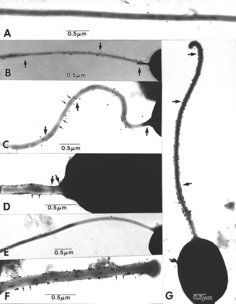
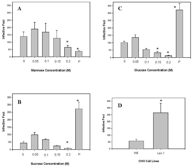
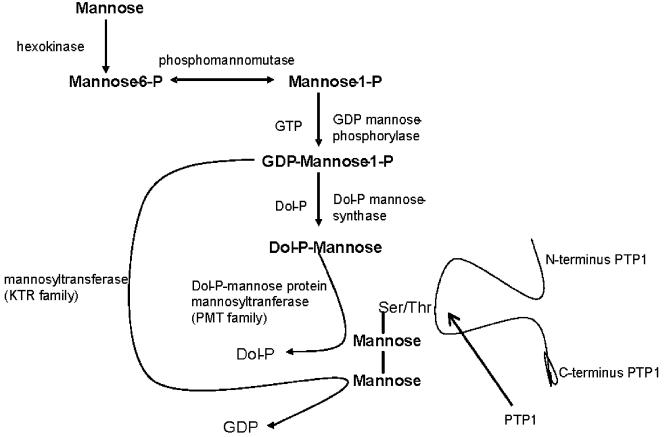
The microsporidia are ubiquitous, obligate intracellular eukaryotic spore-forming parasites infecting a wide range of invertebrates and vertebrates, including humans. The defining structure of microsporidia is the polar tube, which forms a hollow tube through which the sporoplasm is transferred to the host cell. Research on the molecular and cellular biology of the polar tube has resulted in the identification of three polar tube proteins: PTP1, PTP2, and PTP3. The major polar tube protein, PTP1, accounts for at least 70% of the mass of the polar tube. In the present study, PTP1 was found to be posttranslationally modified. Concanavalin A (ConA) bound to PTP1 and to the polar tube of several different microsporidia species. Analysis of the glycosylation of Encephalitozoon hellem PTP1 suggested that it is modified by O-linked mannosylation, and ConA binds to these O-linked mannose residues. Mannose pretreatment of RK13 host cells decreased their infection by E. hellem, consistent with an interaction between the mannosylation of PTP1 and some unknown host cell mannose-binding molecule. A CHO cell line (Lec1) that is unable to synthesize complex-type N-linked oligosaccharides had an increased susceptibility to E. hellem infection compared to wild-type CHO cells. These data suggest that the O-mannosylation of PTP1 may have functional significance for the ability of microsporidia to invade their host cells.


Figures

FIG. 1.
FIG. 1.
Analysis and purification of the major PTP of E. hellem (EhPTP1) by HPLC. This figure demonstrates the typical major PTP peak (EhPTP1) seen with HPLC purification of DTT-solubilized E. hellem PTPs (12-15). The major peak at 35 min in the figure is EhPTP1. (Insert) Lane 1, SDS-PAGE of peak at 35 min stained with SYPRO Ruby; lane 2; immunoblot of 35-min peak using rabbit antibody to recEhPTP1.
FIG. 2.
FIG. 2.
Interaction of EhPTP1 with the lectin ConA. (A) ConA overlay. A DTT-solubilized polar tube preparation was used for SDS-PAGE, transferred to nitrocellulose, and incubated with labeled ConA in the presence and absence of α-methyl-mannopyranoside. ConA bound to a single band (lane 1), and this binding was inhibited by α-methyl-mannopyranoside (lane 2). (B) ConA affinity chromatography. Soluble E. hellem PTPs were loaded onto a ConA-Sepharose column, and specifically retained proteins were eluted with 0.2 M α-methyl-mannopyranoside-0.15 mM NaCl. The flowthrough of the column (e.g., soluble PTPs after loading on the column) and the specific eluate were analyzed by SDS-PAGE followed by immunoblot analysis with rabbit antibody to recEhPTP1. The specific eluate contained EhPTP1 (lane 1), while the flowthrough no longer contained EhPTP1 (lane 2).
FIG. 3.
FIG. 3.
PTP1 reactivity with ConA is seen in multiple microsporidia. Solubilized (2% DTT) polar tube preparations of five microsporidia species, G. americanus (Ga), E. hellem (Eh), E. cuniculi (Ec), E. intestinalis (Ei), and B. algerae (Ba), were used for a lectin overlay with labeled ConA. ConA reacted with a band in each polar tube preparation that had a size consistent with that seen on SDS-PAGE and immunoblot analysis with antibody to PTP1 (12-15).
FIG. 4.
FIG. 4.
Analysis of N-glycosylation of the major PTP of E. hellem (EhPTP1). (A) N-deglycosylation assay results. Coomassie-stained SDS-PAGE gels of EhPTP1 (lanes 1 to 4) and fetuin (lanes 5 to 8) were treated with NANase II, O-glycosidase, and/or PHGase F. Lanes 1 and 5, no enzymes; lanes 2 and 6, treatment with NANase II; lanes 3 and 7, treatment with NANase II and O-glycosidase; lanes 4 and 8, treatment with NANase II, O-glycosidase, and PNGase F. There was no apparent molecular weight change of EhPTP1 with treatment. Fetuin was used as control and demonstrated the expected changes in molecular weight following enzymatic treatment. (B) ConA reactivity of EhPTP1 following N-deglycosylation. Shown are the results of ConA lectin overlay of EhPTP1 without treatment (lane 1), EhPTP1 treated with NANase II (lane 2), EhPTP1 treated with NANase II and O-glycosidase (lane 3), and EhPTP1 treated with NANase II, O-glycosidase, and PNGase F (lane 4). There was no significant change in the molecular weight of EhPTP1 with treatment. ConA still bound to EhPTP1 after treatment with NANase II, O-glycosidase, and PNGase F.
FIG. 5.
FIG. 5.
Analysis of O-glycosylation of EhPTP1. HPLC-purified EhPTP1 was treated with 0.1 N NaOH at 50°C for 0, 10, 20, 30, 40, and 50 min. After treatment, samples were analyzed by SDS-PAGE followed by either labeled ConA overlay (top panel) or immunoblotting with polyclonal rabbit antibody to HPLC-purified nEhPTP1 (bottom panel). The intensities of all bands were determined by densitometry. The numbers on each lane reflect the band intensity compared to the amount of binding present at time zero (100%). At 40 min, EhPTP1 was still present by immunoblotting, but there was no ConA binding.
FIG. 6.
FIG. 6.
Electron micrographs of the reaction of extruded microsporidian polar tubes of E. hellem (A to D) and B. algerae (E to G) with colloidal gold-labeled ConA. (A) Fully extruded E. hellem polar tube demonstrating no reaction with colloidal gold-labeled ConA in the presence of 0.2 M α-mannopyranoside. A few unbound gold particles are present in the background. (B) Fully extruded E. hellem polar tube decorated with small clusters of gold particles (arrows). Note the intense clustering on the portion of the tube near the spore. (C) Fully extruded E. hellem polar tube decorated with numerous individual particles of gold (arrows). The periodicity of the polar tube substructure is evident (small arrows). (D) Partially extruded E. hellem polar tube decorated with a few individual particles of gold (arrows). The localization appears to be more intense in the area close to the spore. At this relatively early stage of extrusion, the polar tube substructure is evident (small arrows). (E) Fully extruded B. algerae polar tube demonstrating no reaction with colloidal gold-labeled ConA in the presence of 0.2 M α-mannopyranoside. (F) Partially extruded B. algerae polar tube, uniformly decorated with many particles of gold along its surface (arrows). The periodicity of the polar tube substructure is evident (small arrows). (G) Fully extruded B. algerae polar tube intensely decorated with individual and clusters of gold particles along its entire length (arrows). While ConA binding was present on polar tubes of both E. hellem and B. algerae, the binding appeared to be significantly more abundant on Brachiola polar tubes.
FIG. 7.
FIG. 7.
Studies on the effects of carbohydrates and host cell glycosylation on the infection of host cells by E. hellem. The ability of E. hellem to infect RK13 cells in the presence of different concentrations of carbohydrates and the effects of pretreatment of host cells with carbohydrates for 3 days preceding infection were assessed. Each well was infected with 104 E. hellem spores, and infection was quantified as the number of infected foci seen under a fluorescence microscope in 20 40× fields per well. Comparisons of infective foci counts were done with Student's t test. (A) Mannose results. RK13 cells were cultured with various concentrations of α-mannose (0, 0.05, 0.1, 0.15, and 0.2 M) at the time of infection. In addition, RK13 cells were pretreated (P) with 0.1 M mannose for 3 days prior to infection and then infected and cultured in the absence of mannose. Significant inhibition of infection was seen with either 0.2 M α-mannose in the medium during infection or pretreatment of cells with 0.1 M α-mannose prior to infection (P < 0.05). (B) Sucrose results. RK13 cells were cultured with various concentrations of sucrose (0, 0.05, 0.1, 0.15, and 0.2 M) at the time of infection. In addition, RK13 cells were pretreated (P) with 0.1 M sucrose for 3 days prior to infection and then infected and cultured in the absence of sucrose. Significant inhibition of infection was seen with 0.2 M sucrose in the medium during infection. A significant increase in infection was seen with pretreatment of cells with 0.1 M sucrose prior to infection (P < 0.05). Infection was also increased by the presence of 0.05 M sucrose in the medium. (C) Glucose results. RK13 cells were cultured with various concentrations of glucose (0, 0.05, 0.1, 0.15, and 0.2 M) at the time of infection. In addition, RK13 cells were pretreated (P) with 0.1 M glucose for 3 days prior to infection and then infected and cultured in the absence of glucose. Significant inhibition of infection was seen with 0.15 and 0.2 M glucose in the medium during infection. A significant increase in infection was seen with pretreatment of cells with 0.1 M glucose prior to infection (P < 0.05). Infection was also increased by the presence of 0.05 M glucose in the medium. (D) CHO cell results. The infectivity of E. hellem for wild-type CHO cells (W5) and mutant CHO cells (Lec1), which could bind more ConA than wild type, was assessed. Using the same infective dose of E. hellem, the number of infective foci was significantly higher in Lec1 cells than in W5 (wild-type) CHO cells (P < 0.05).
FIG. 8.
FIG. 8.
A proposed O-mannosylation pathway for E. cuniculi. Analysis of the published E. cuniculi genome (10, 36) demonstrated the presence of the enzymes required for the O-mannosylation of proteins.

References

    1. Aronson, M., O. Medalia, L. Schori, D. Mirelman, N. Sharon, and I. Ofek. 1979. Prevention of colonization of the urinary tract of mice with Escherichia coli by blocking of bacterial adherence with methyl alpha-D-mannopyranoside. J. Infect. Dis. 139:329-332. - PubMed
    1. Cali, A., L. M. Weiss, and P. M. Takvorian. 2002. Brachiola algerae spore membrane systems, their activity during extrusion, and a new structural entity, the multilayered interlaced network, associated with the polar tube and the sporoplasm. J. Eukaryot. Microbiol. 49:164-174. - PubMed
    1. Chen, X. M., and N. F. LaRusso. 2000. Mechanisms of attachment and internalization of Cryptosporidium parvum to biliary and intestinal epithelial cells. Gastroenterology 118:368-379. - PubMed
    1. Delbac, F., I. Peuvel, G. Metenier, E. Peyretaillade, and C. P. Vivares. 2001. Microsporidian invasion apparatus: identification of a novel polar tube protein and evidence for clustering of ptp1 and ptp2 genes in three Encephalitozoon species. Infect. Immun. 69:1016-1024. - PMC - PubMed
    1. Delbac, F., P. Peyret, G. Metenier, D. David, A. Danchin, and C. P. Vivares. 1998. On proteins of the microsporidian invasive apparatus: complete sequence of a polar tube protein of Encephalitozoon cuniculi. Mol. Microbiol. 29:825-834. - PubMed

Publication types

LinkOut - more resources