

Cytokine response to infection with Bacillus anthracis spores
- PMID: 15501768
- PMCID: PMC523056
- DOI: 10.1128/IAI.72.11.6382-6389.2004
Cytokine response to infection with Bacillus anthracis spores
Abstract
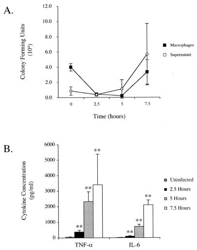
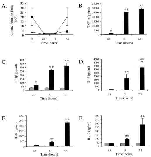
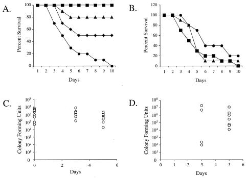
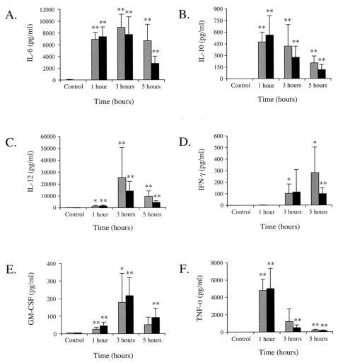
Bacillus anthracis, the etiological agent of anthrax, is a gram-positive, spore-forming bacterium. The inhalational form of anthrax is the most severe and is associated with rapid progression of the disease and the outcome is frequently fatal. Transfer from the respiratory epithelium to regional lymph nodes appears to be an essential early step in the establishment of infection. This transfer is believed to occur by means of carriage within alveolar macrophages following phagocytosis. Therefore, the ability of B. anthracis to transit through the host macrophage or dendritic cell appears to be an early and critical step in B. anthracis pathogenesis. In this work, we examined the cytokine responses to spore infection in mouse primary peritoneal macrophages, in primary human dendritic cells, and during a spore aerosol infection model utilizing the susceptible A/J mouse strain. We demonstrated that both mouse peritoneal macrophages and human dendritic cells exhibited significant intracellular bactericidal activity during the first hours following uptake, providing the necessary time to mount a cytokine response prior to cell lysis. Strong tumor necrosis factor (TNF-alpha) and interleukin-6 (IL-6) responses were seen in mouse peritoneal macrophages. In addition to TNF-alpha and IL-6, human dendritic cells produced the cytokines IL-1beta, IL-8, and IL-12. A mixture of Th1 and Th2 cytokines were detected in sera obtained from infected animals. In this study, we provide further evidence of an acute cytokine response when cells in culture and mice are infected with B. anthracis spores.
Figures
Similar articles
-
Transport of Bacillus anthracis from the lungs to the draining lymph nodes is a rapid process facilitated by CD11c+ cells.Microb Pathog. 2010 Jul-Aug;49(1-2):38-46. doi: 10.1016/j.micpath.2010.02.004. Epub 2010 Feb 25. Microb Pathog. 2010. PMID: 20188814
-
Modulation of cytokine production and enhancement of cell viability by TLR7 and TLR9 ligands during anthrax infection of macrophages.FEMS Immunol Med Microbiol. 2006 Aug;47(3):369-79. doi: 10.1111/j.1574-695X.2006.00096.x. FEMS Immunol Med Microbiol. 2006. PMID: 16872373
-
The use of a model of in vivo macrophage depletion to study the role of macrophages during infection with Bacillus anthracis spores.Microb Pathog. 2004 Oct;37(4):169-75. doi: 10.1016/j.micpath.2004.06.013. Microb Pathog. 2004. PMID: 15458777
-
New aspects of the infection mechanisms of Bacillus anthracis.Ann Agric Environ Med. 2012;19(4):613-8. Ann Agric Environ Med. 2012. PMID: 23311776 Review.
-
A review of the interaction of Bacillus anthracis with cells of the innate immune response.Berl Munch Tierarztl Wochenschr. 2006 May-Jun;119(5-6):216-21. Berl Munch Tierarztl Wochenschr. 2006. PMID: 16729468 Review.
Cited by
-
Activity of the Bacillus anthracis 20 kDa protective antigen component.BMC Infect Dis. 2008 Sep 22;8:124. doi: 10.1186/1471-2334-8-124. BMC Infect Dis. 2008. PMID: 18808698 Free PMC article.
-
Cytokine response and survival of mice immunized with an adenovirus expressing Bacillus anthracis protective antigen domain 4.Infect Immun. 2006 Feb;74(2):1009-15. doi: 10.1128/IAI.74.2.1009-1015.2006. Infect Immun. 2006. PMID: 16428747 Free PMC article.
-
Inter-α inhibitor proteins: a novel therapeutic strategy for experimental anthrax infection.Shock. 2011 Jan;35(1):42-4. doi: 10.1097/SHK.0b013e3181e83204. Shock. 2011. PMID: 20523269 Free PMC article.
-
New insights into gastrointestinal anthrax infection.Trends Mol Med. 2015 Mar;21(3):154-63. doi: 10.1016/j.molmed.2014.12.003. Epub 2014 Dec 19. Trends Mol Med. 2015. PMID: 25577136 Free PMC article. Review.
-
Transcriptional and apoptotic responses of THP-1 cells to challenge with toxigenic, and non-toxigenic Bacillus anthracis.BMC Immunol. 2008 Nov 13;9:67. doi: 10.1186/1471-2172-9-67. BMC Immunol. 2008. PMID: 19014542 Free PMC article.
References
-
- Agrawal, A., J. Lingappa, S. H. Leppla, S. Agrawal, A. Jabbar, C. Quinn, and B. Pulendran. 2003. Impairment of dendritic cells and adaptive immunity by anthrax lethal toxin. Nature 424:329-334. - PubMed
-
- Barnes, J. M. 1947. The development of anthrax following administration of spores by inhalation. Br. J. Exp. Pathol. 28:385-394.
-
- Dalldorf, F. G., A. F. Kaufmann, and P. S. Brachman. 1971. Woolsorter's disease. Arch. Pathol. 92:418-426. - PubMed
-
- Dang, O., L. Navarro, K. Anderson, and M. David. 2004. Cutting edge: anthrax lethal toxin inhibits activation of IFN-regulatory factor 3 by lipopolysaccharide. J. Immunol. 172:747-751. - PubMed
-
- Dixon, T. C., A. A. Fadl, T. M. Koehler, J. A. Swanson, and P. C. Hanna. 2000. Early Bacillus anthracis-macrophage interactions: intracellular survival and escape. Cell Microbiol. 2:453-463. - PubMed
MeSH terms
Substances
LinkOut - more resources
Full Text Sources
Other Literature Sources
Medical
