

doi: 10.1128/IAI.72.11.6680-6684.2004.
Shiga toxin binding to isolated porcine tissues and peripheral blood leukocytes
Affiliations
- PMID: 15501802
- PMCID: PMC523021
- DOI: 10.1128/IAI.72.11.6680-6684.2004
Item in Clipboard
Shiga toxin binding to isolated porcine tissues and peripheral blood leukocytes
Infect Immun.
2004 Nov.
Abstract
Shiga toxin (Stx) binding sites in porcine tissues and leukocytes were identified by the use of Stx overlay and anti-CD77/Gb3 immunoassays. Stx1 and Stx2 bound to similar tissue locations and leukocytes, although some differences were noted. Previously unreported Stx binding sites were identified in kidney tubules, intestinal lymphoid aggregates, sinusoidal liver cells, alveolar macrophages, and peripheral blood leukocytes.
Figures
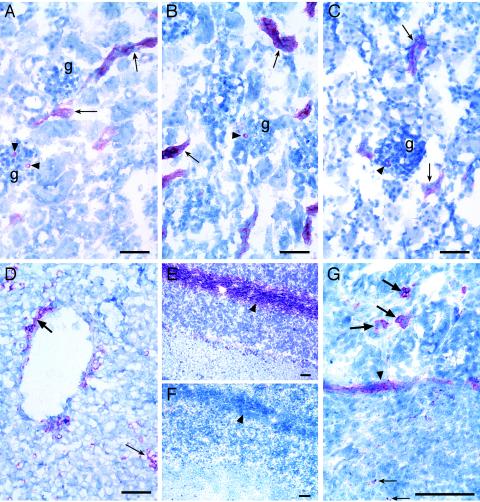
Immunohistochemical identification of Stx binding sites and Gb3 in neonatal porcine tissues. Anti-CD77/Gb3 (A), Stx1 (B), and Stx2 (C) binding to tubules (arrows) and glomeruli (arrowheads) in sections of the kidney (g, glomerulus) are shown. (D) Stx2 binding to endothelial cells lining a vessel (thick arrow) and sinusoidal cells within the parenchyma (thin arrow) of the liver. Anti-CD77/Gb3 (E), but not Stx1 (F), binding to nerve fibrils in the white matter (arrowheads) of the cerebellum was observed. (G) Stx2 binding within a Peyer's patch (thin arrow), to smooth muscle (arrowhead), and to cells within the villous lamina propria (thick arrows) in the ileum. Digital images were captured directly with a Digital Spot RT Slider camera (Diagnostic Instruments, Inc., Sterling Heights, Mich.) using MetaVue, version 5.0.7, imaging software (Universal Imaging Corp., Downingtown, Pa.). Bar = 50 μm.
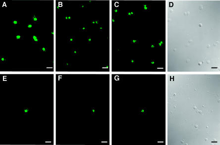
Indirect immunofluorescent detection of Stx binding and Gb3 on porcine peripheral blood leukocytes. Anti-CD77/Gb3 (A), Stx1 (B), and Stx2 (C) binding to cells on granulocyte smears are shown. Anti-CD77/Gb3 (E), Stx1 (F), and Stx2 (G) binding to cells on mononuclear leukocyte smears are also shown. (D and H) Differential interference contrast illumination of the same fields as those in panels C and G, respectively. Images were obtained as described in the legend to Fig. 1. Bar = 25 μm.
References
-
- Barnes, D., S. Aggarwal, S. Thomsen, M. Fitzmaurice, and R. Richards-Kortum. 1993. A characterization of the fluorescent properties of circulating human eosinophils. Photochem. Photobiol. 58:297-303. - PubMed
-
- Boyd, B., G. Tyrrell, M. Maloney, C. Gyles, J. Brunton, and C. Lingwood. 1993. Alteration of the glycolipid binding specificity of the pig edema toxin from globotetraosyl to globotriaosyl ceramide alters in vivo tissue targeting and results in a verotoxin 1-like disease in pigs. J. Exp. Med. 177:1745-1753. - PMC - PubMed
-
- Chark, D., A. Nutikka, N. Trusevych, J. Kuzmina, and C. Lingwood. 2004. Differential carbohydrate epitope recognition of globotriaosyl ceramide by verotoxins and a monoclonal antibody. Eur. J. Biochem. 271:405-417. - PubMed
MeSH terms
Substances
LinkOut - more resources
Full Text Sources
