. 2005 Jan;88(1):680-9.
doi: 10.1529/biophysj.104.048025. Epub 2004 Oct 22.

Mechanical properties of Xenopus egg cytoplasmic extracts

Affiliations

Mechanical properties of Xenopus egg cytoplasmic extracts

M T Valentine et al. Biophys J. 2005 Jan.

Abstract

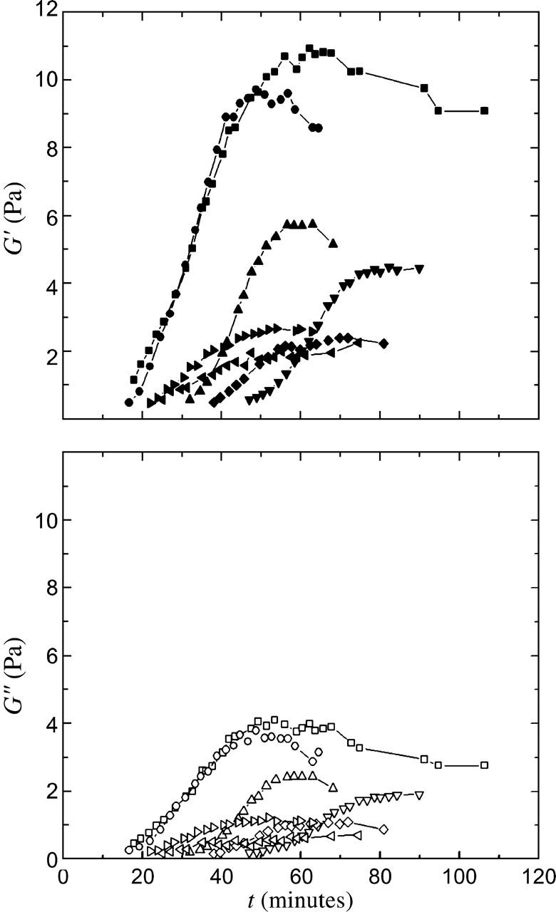
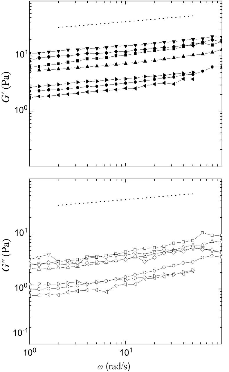
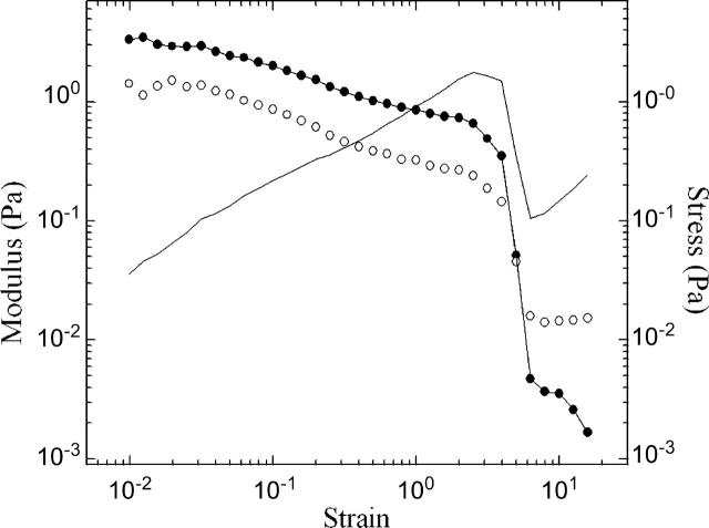
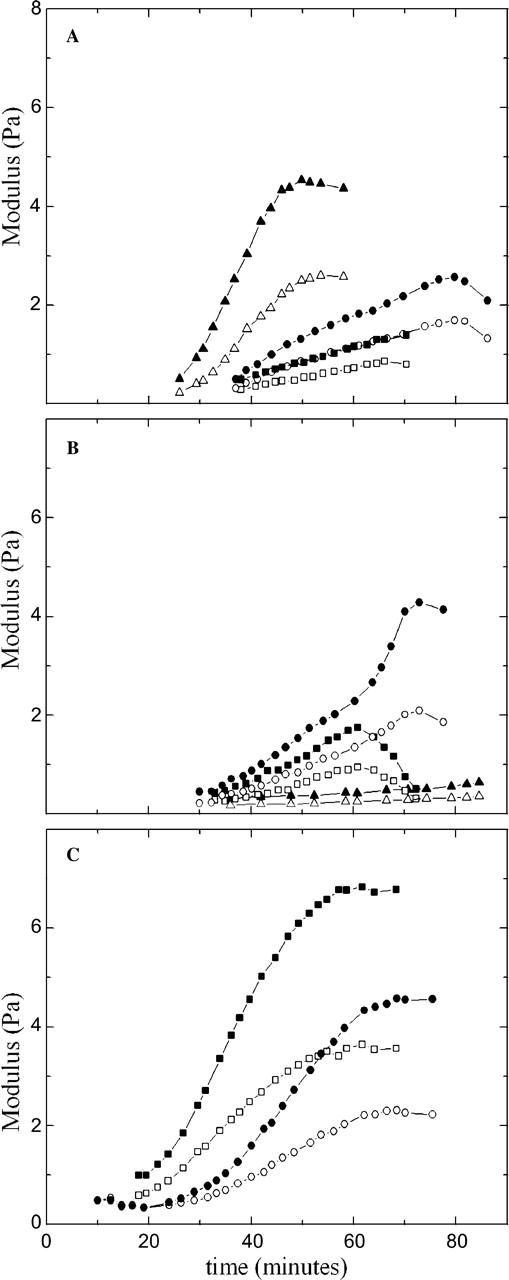
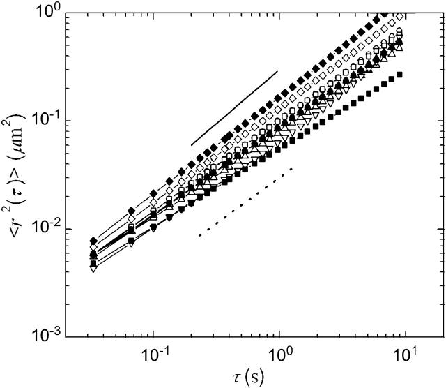
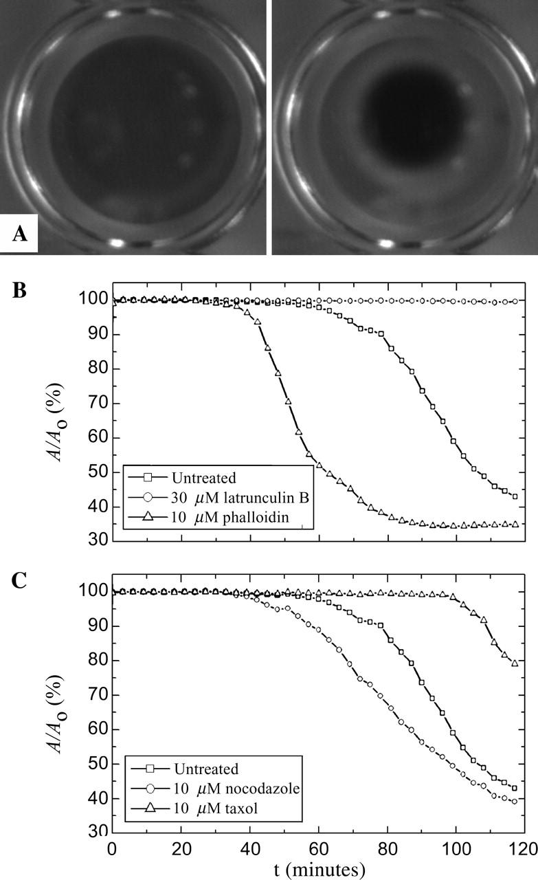
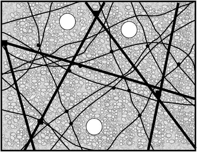
Cytoplasmic extracts prepared from Xenopus laevis eggs are used for the reconstitution of a wide range of processes in cell biology, and offer a unique environment in which to investigate the role of cytoplasmic mechanics without the complication of preorganized cellular structures. As a step toward understanding the mechanical properties of this system, we have characterized the rheology of crude interphase extracts. At macroscopic length scales, the extract forms a soft viscoelastic solid. Using a conventional mechanical rheometer, we measure the elastic modulus to be in the range of 2-10 Pa, and loss modulus in the range of 0.5-5 Pa. Using pharmacological and immunological disruption methods, we establish that actin filaments and microtubules cooperate to give mechanical strength, whereas the intermediate filament cytokeratin does not contribute to viscoelasticity. At microscopic length scales smaller than the average network mesh size, the response is predominantly viscous. We use multiple particle tracking methods to measure the thermal fluctuations of 1 microm embedded tracer particles, and measure the viscosity to be approximately 20 mPa-s. We explore the impact of rheology on actin-dependent cytoplasmic contraction, and find that although microtubules modulate contractile forces in vitro, their interactions are not purely mechanical.


Figures

FIGURE 1
FIGURE 1
Time-evolution of the viscoelastic response of untreated cytoplasmic extracts, with measurements taken every 2–3 min with ω = 1 rad/s and γo = 0.05. Each curve represents an independent measurement, showing the variation in the response. In all cases, the extracts form weak viscoelastic solids, with elastic moduli G′ (top, solid symbols) in the range of 2–10 Pa, and viscous moduli G″ (bottom, open symbols) in the range of 0.5–5 Pa.
FIGURE 2
FIGURE 2
Frequency-dependence of the viscoelastic response of untreated cytoplasmic extracts, with γo = 0.05. Each curve represents an independent measurement, showing the variation in the response. In all cases, the elastic modulus dominates the loss modulus, and both G′ (top, solid symbols) and G″ (bottom, open symbols) show a weak dependence on frequency. The dotted line has a slope of 0.15, indicating that at these frequencies the extract responds as a viscoelastic solid.
FIGURE 3
FIGURE 3
Representative data showing the strain-dependence of the stress (line), G′ (solid symbols), and G″ (open symbols), obtained with a constant frequency ω = 1 rad/s. The linear regime is small, with strain softening occurring for γo > 0.01. G′ dominates until very large strains. At the highest strains, G″ prevails; we cautiously estimate the viscosity η = ω−1 G″ ∼ 20 mPa-s.
FIGURE 4
FIGURE 4
Storage (solid symbols) and loss (open symbols) moduli as a function of time after warming for extracts treated with (A) 10 μM phalloidin, (B) 10 μM taxol, and (C) 500 nM taxol. In each case, the extracts form weak viscoelastic solids, similar to the untreated native cytoplasmic gels. The time dependence of G′ and G″ is similar, and G″/G′ is ∼0.5.
FIGURE 5
FIGURE 5
Ensemble-averaged MSDs of particles moving in untreated extract (□) as well as extracts that have been treated with 30 μM latrunculin B (○), 10 μM nocodazole (▿), 10 μM taxol (⋄), and 500 nM taxol (▵), after a 10 min (open symbols) or 30 min incubation (solid symbols) at room temperature. Our data evolve slightly subdiffusively with lag time, indicating that the cytosol is not a pure fluid on these length scales. The solid line represents a slope of 1, as expected for a pure viscous fluid, and the dotted line a slope of 0.85. Although not a simple fluid, the material is predominantly viscous with viscosity in the range of 10–30 mPa-s. We measure no significant change in particle dynamics upon disassembly of either the actin or microtubule networks, or the stabilization of the microtubule network, and observe no significant changes in viscosity for waiting times of up to 30 min. At 30 min, the extracts are still dominated by viscous relaxation, with viscosity in the range of 10–30 mPa-s.
FIGURE 6
FIGURE 6
(A) Time-lapsed pictures demonstrating gelation/contraction for untreated extracts at t = 0 and t = 120 min. (B) Effect of actin depolymerizing and stabilizing drugs on contraction, here represented by the contracted gel area A normalized by initial gel area Ao. The disassembly of the actin network by latrunculin B prevents contraction, whereas addition of the stabilizing agent phalloidin accelerates the onset of contraction. (C) Effect of microtubule depolymerizing and stabilizing drugs on contraction. The disruption of the microtubule network by nocodazole accelerates whereas taxol stabilization inhibits contraction.
FIGURE 7
FIGURE 7
Sketch of the mechanically important structures in egg extract. F-actin, microtubules, and cytokeratin form a composite network stiffened by numerous cross-linking proteins (solid circles and squares). Protein-resistant micron-sized colloidal particles (white circles) do not interact with the elastic mesh; rather, they move within mechanically distinct microenvironments. The cytoskeleton is bathed in a concentrated protein solution, the cytosol, represented by the small macromolecules (grayscale, in background). This concentrated suspension increases the microscopic viscosity to ∼20 times that of water.

Similar articles

Cited by

References

    1. Alberts, B., A. Johnson, J. Lewis, M. Raff, K. Roberts, and P. Walter. 2002. Molecular Biology of the Cell. Garland, New York.
    1. Andra, K., H. Lassman, R. Bittner, S. Shomy, R. Fassler, R. Propst, and G. Wiche. 1997. Targeted inactivation of plectin reveals essential function in maintaining the integrity of skin, muscle, and heart cytoarchitecture. Genes Dev. 11:3143–3156. - PMC - PubMed
    1. Bausch, A. R., W. Möller, and E. Sackmann. 1999. Measurement of local viscoelasticity and forces in living cells by magnetic tweezers. Biophys. J. 76:573–579. - PMC - PubMed
    1. Benink, H. A., C. A. Mandato, and W. M. Bement. 2000. Analysis of cortical flow models in vivo. Mol. Biol. Cell. 11:2553–2563. - PMC - PubMed
    1. Boal, D. 2002. Mechanics of the Cell. Cambridge University Press, Cambridge.

Publication types