. 2005 Jan;88(1):603-8.
doi: 10.1529/biophysj.104.049312. Epub 2004 Oct 22.

The high-resolution architecture and structural dynamics of Bacillus spores

Affiliations

The high-resolution architecture and structural dynamics of Bacillus spores

Marco Plomp et al. Biophys J. 2005 Jan.

Abstract

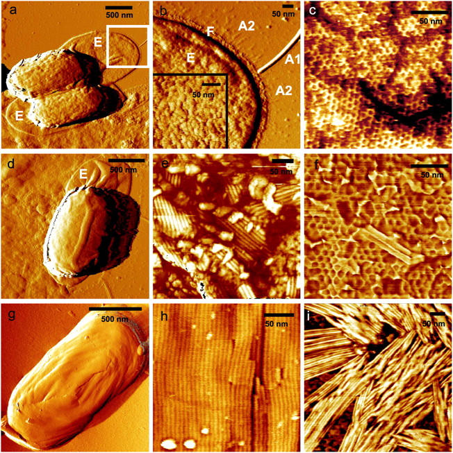
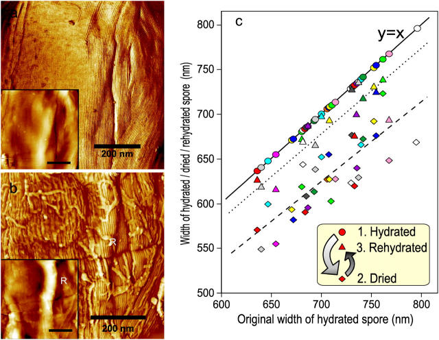
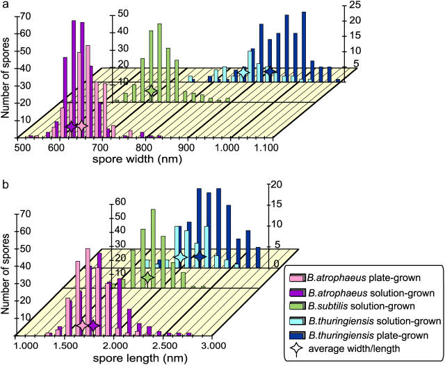
The capability to image single microbial cell surfaces at nanometer scale under native conditions would profoundly impact mechanistic and structural studies of pathogenesis, immunobiology, environmental resistance, and biotransformation. Here, using in vitro atomic force microscopy, we have directly visualized high-resolution native structures of bacterial endospores, including the exosporium and spore coats of four Bacillus species in air and water environments. Our results demonstrate that the mechanisms of spore coat self-assembly are similar to those described for inorganic and macromolecular crystallization. The dimensions of individual Bacillus atrophaeus spores decrease reversibly by 12% in response to a change in the environment from fully hydrated to air-dried state, establishing that the dormant spore is a dynamic physical structure. The interspecies distributions of spore length and width were determined for four species of Bacillus spores in water and air environments. The dimensions of individual spores differ significantly depending upon species, growth regimes, and environmental conditions. These findings may be useful in the reconstruction of environmental and physiological conditions during spore formation and for modeling the inhalation and dispersal of spores. This study provides a direct insight into molecular architecture and structural variability of bacterial endospores as a function of spatial and developmental organizational scales.


Figures

FIGURE 1
FIGURE 1
The spore coats of B. thuringiensis (ac), B. cereus (df), and B. atrophaeus (g and h) consist of crystalline layers of honeycomb and rodlet structures. B. thuringiensis (a) and B. cereus (d) spores are surrounded by an exosporium (E), which collapses and adheres to the substrate when spores are air-dried. The exosporium (b) is 25–40-nm thick and has a ∼3-nm thick and 20–30-nm wide footstep (F) with numerous thin (∼1 nm) and short (50–60 nm) hair-like appendages (A2) attached to it. Additionally, there are typically 4–8 tubular appendages (A1) attached to the exosporium with diameters and lengths of 3–12 nm and 300–1200 nm, respectively. Underneath the exosporium, a honeycomb crystalline layer defines the outer surface of B. thuringiensis spores (c). This honeycomb layer is formed by domains with random crystalline orientations that are separated by linear defects. In e and h, the crystalline rodlet structures of B. cereus and B. atrophaeus spore coats are shown. B. cereus spores contain a crystalline honeycomb structure (f) beneath the exterior rodlet layer. B. thuringiensis spore coats do not contain rodlet structures. Rodlet assemblies can be seen adsorbed to the substrate (i). Scale bar, 500 nm in a, d, and g; 50 nm in b, c, e, f, h, and i.
FIGURE 2
FIGURE 2
The effects of changing the B. atrophaeus spore environment from hydrated to dehydrated states. (a) Phase image and height image (inset) detail of a B. atrophaeus spore in water, showing several shallow wrinkles. (b) The same spore after drying, showing many adsorbed stray rodlets, and a 60-nm high ridge (indicated with R). Loose stray rodlets sediment from the bulk solution upon drying of the sample. Stray rodlets are present on the surfaces of all air-dried spores. (c) Width change of 35 individual B. atrophaeus spores. Depicted is the individual measured spore width for a set of dehydrated (24 h) (diamonds, dashed trend line) and rehydrated (2 h) spores (triangles, dotted trend line), as a function of the size of the originally hydrated spore. For ease of comparison, the original hydrated spore width is (redundantly) depicted as circles, which by definition lie on the solid y = x line. Thus, the three data points for one individual spore, depicted with the same color, are all on the same vertical line. Several spores detached from the substrate during rehydration, which resulted in fewer experimental points for rehydrated spores (triangles). On average, spore size is reduced to 88% for dried spores, and returns to 97% of the original width for rehydrated spores (see text). Scale bar in a and b, 200 nm.
FIGURE 3
FIGURE 3
Distribution of spore width (a) and length (b) for plate-grown (pg) and solution-grown (sg) B. atrophaeus and B. thuringiensis spores, and solution-grown B. subtilis spores. Average width and length are indicated with stars. Statistics for these distributions are given in Table 1.

References

    1. Aronson, A. I., and P. Fitz-James. 1976. Structure and morphogenesis of the bacterial spore coat. Bacteriol. Rev. 40:360–402. - PMC - PubMed
    1. Binnig, G., C. F. Quate, and C. Gerber. 1986. Atomic force microscope. Phys. Rev. Lett. 56:930–933. - PubMed
    1. Bustamante, C., C. Rivetti, and D. Keller. 1997. Scanning force microscopy under aqueous solutions. Curr. Opin. Struct. Biol. 7:709–716. - PubMed
    1. Chada, V. G. R., E. A. Sanstad, R. Wang, and A. Driks. 2003. Morphogenesis of Bacillus spore surfaces. J. Bacteriol. 185:6255–6261. - PMC - PubMed
    1. Chernov, A. A. 1984. Modern Crystallography III. Crystal Growth. Springer-Verlag, Berlin.

Publication types

LinkOut - more resources