

Selective lumbar nerve root blocks with CT fluoroscopic guidance: technique, results, procedure time, and radiation dose
Affiliations
- PMID: 15502144
- PMCID: PMC7976407
Item in Clipboard
Selective lumbar nerve root blocks with CT fluoroscopic guidance: technique, results, procedure time, and radiation dose
AJNR Am J Neuroradiol.
2004 Oct.
Abstract
CT fluoroscopy may be used as a rapid and effective means of guiding needle placement when performing selective lumbar nerve root blocks. In this set of patients, the average external radiation dose was 0.73 mrem per procedure, with an average of 2 seconds of CT-fluoroscopy time and four images per procedure. Average physician room time was 7 minutes. Use of intermittent CT fluoroscopy during lumbar selective nerve root blocks can result in minimal radiation dose levels and procedure times that are comparable to fluoroscopic guidance.
Copyright American Society of Neuroradiology
Figures
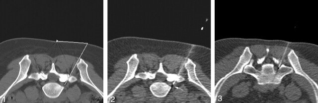
Initial scout image shows the nerve root in the neural foramen, indicating an appropriate injection level. The distance tool is used to calculate an appropriate skin injection site. Once the technologists become familiar with the anatomy, many studies can be done with only one or two scout images.
Image from CT fluoroscopy-guided nerve root block demonstrates the needle tip in the outer neural foramen. Note that the image is not as sharp as standard CT images due to the low mA used with this technique.
S1 nerve root block shows the needle traversing the sacral foramen and lying directly adjacent to the S1 nerve.
References
-
- Murtagh R. The neuroradiologist as pain therapist. AJNR Am J Neuroradiol 1998;19:353–354
-
- Quinn SF, Murtagh R. CT-guided nerve root sleeve block and ablation. AJR Am J Roentgenol 1988;151:1213–1216 - PubMed
-
- Murtagh R. The art and science of nerve root and facet blocks. Neuroimaging Clin N Am 2000;10:465–477 - PubMed
-
- Katada K, Anno H, Ogura Y, et al. Early clinical experience with real-time CT fluoroscopy. Nippon Acta Radiol 1994;54:1172–1174 - PubMed
MeSH terms
Substances
LinkOut - more resources
Full Text Sources
Medical