. 2004 Nov;2(11):e333.
doi: 10.1371/journal.pbio.0020333. Epub 2004 Oct 5.

Protein thiol modifications visualized in vivo

Affiliations

Protein thiol modifications visualized in vivo

Lars I Leichert et al. PLoS Biol. 2004 Nov.

Abstract

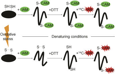
Thiol-disulfide interconversions play a crucial role in the chemistry of biological systems. They participate in the major systems that control the cellular redox potential and prevent oxidative damage. In addition, thiol-disulfide exchange reactions serve as molecular switches in a growing number of redox-regulated proteins. We developed a differential thiol-trapping technique combined with two-dimensional gel analysis, which in combination with genetic studies, allowed us to obtain a snapshot of the in vivo thiol status of cellular proteins. We determined the redox potential of protein thiols in vivo, identified and dissected the in vivo substrate proteins of the major cellular thiol-disulfide oxidoreductases, and discovered proteins that undergo thiol modifications during oxidative stress. Under normal growth conditions most cytosolic proteins had reduced cysteines, confirming existing dogmas. Among the few partly oxidized cytosolic proteins that we detected were proteins that are known to form disulfide bond intermediates transiently during their catalytic cycle (e.g., dihydrolipoyl transacetylase and lipoamide dehydrogenase). Most proteins with highly oxidized thiols were periplasmic proteins and were found to be in vivo substrates of the disulfide-bond-forming protein DsbA. We discovered a substantial number of redox-sensitive cytoplasmic proteins, whose thiol groups were significantly oxidized in strains lacking thioredoxin A. These included detoxifying enzymes as well as many metabolic enzymes with active-site cysteines that were not known to be substrates for thioredoxin. H(2)O(2)-induced oxidative stress resulted in the specific oxidation of thiols of proteins involved in detoxification of H(2)O(2) and of enzymes of cofactor and amino acid biosynthesis pathways such as thiolperoxidase, GTP-cyclohydrolase I, and the cobalamin-independent methionine synthase MetE. Remarkably, a number of these proteins were previously or are now shown to be redox regulated.


Conflict of interest statement

The authors have declared that no conflicts of interest exist.

Figures

Figure 1
Figure 1. Schematic Overview of Our Differential Thiol-Trapping Technique
Under normal growth conditions (top), a hypothetical cytoplasmic protein present within the complex mixture of the crude whole-cell extract is fully reduced. Upon incubation in TCA, all thiol-disulfide exchange reactions are quenched and the cells lyse. In the first thiol-trapping step, the protein is denatured and incubated with IAM. Accessible thiol groups are quickly carbamidomethylated (CAM) and blocked for the subsequent reduction/alkylation steps. After TCA precipitation and washing, DTT is added to reduce oxidized cysteines, and 14C-labeled IAM is used to modify potentially newly released, accessible cysteines. Under oxidative stress conditions (bottom) the cysteine residues become modified (e.g., sulfenic acid and disulfide bonds). In the first trapping step, IAM cannot attack the oxidized disulfide bond. Only after reduction with DTT are the cysteines accessible to the 14C-labeled IAM. Therefore, the 14C radioactivity correlates with the degree of thiol modification in the protein. The differentially trapped protein species are chemically identical regardless of their original thiol-disulfide status. This ensures their identical migration behavior on 2D gel electrophoresis.
Figure 2
Figure 2. Overall Thiol-Disulfide State of Cellular Proteins in Exponentially Growing E. coli Wild-Type Cells
(A) Colored overlay of the Coomassie blue–stained 2D gel (shown in green) and the phosphor image (shown in red) of a differentially trapped protein extract from exponentially growing E. coli wild-type cells. Proteins with a high ratio of 14C activity/protein appear red; proteins with a low ratio appear green. Protein spots with a ratio of 14C activity/protein greater than 2.0 are indicated by an arrow, while circles label abundant proteins without cysteines. (B) Distribution of the 14C activity/protein ratio in the 100 most abundant protein spots found on a Coomassie blue–stained gel. Bars representing spots with a ratio higher than 2.0 are colored red and are labeled with the name of the protein(s) they represent. (C) Distribution of the 14C activity/protein ratio in the 100 most intense protein spots found on the phosphor images. Bars representing spots with a ratio higher than 2.0 are colored red and are labeled with the name of the protein(s) they represent. (D) Regular and reverse trapping of exponentially growing E. coli wild-type cells. Details of colored overlays of stained protein gels (shown in green) and phosphor images (shown in red) of cell extracts upon regular trapping (top) and reverse trapping (bottom).
Figure 3
Figure 3. Identification of In Vivo Substrate Proteins of the Periplasmic Disulfide Bond Oxidase DsbA
(A) Colored overlay of the stained 2D gel (shown in green) and the phosphor image (shown in red) of differentially trapped protein extract from exponentially growing E. coli wild-type cells. Proteins with a high 14C activity/protein ratio appear red, while proteins with a low ratio appear green. Proteins that were found to have significantly lower ratio of 14C activity/protein in the dsbA::kan strain (B and C) are labeled. (B) Overlay of the stained 2D gel (shown in green) and the phosphor image (shown in red) of a differentially trapped protein extract from exponentially growing dsbA::kan cells. Proteins that were found to have a significantly lower 14C activity/protein ratio in dsbA::kan cells than in wild-type strain (A) are marked with an arrowhead. A circle marks the position of DsbA on the wild-type gel. (C) Overlay of the stained 2D gel (shown in green) and the phosphor image (shown in red) of a differentially trapped protein extract from E. coli dsbA::kan cells growing under oxygen limitation. Arrowheads label proteins that were found to have a significantly lower 14C activity/protein ratio than the wild-type strain grown under oxygen limitation. A circle marks the position of DsbA on the wild-type gel.
Figure 4
Figure 4. Identification of the In Vivo Substrate Proteins of Thioredoxin A
(A) Colored overlay of the stained 2D gel (shown in green) and the phosphor image (shown in red) of differentially trapped protein extract from exponentially growing E. coli wild-type cells. Proteins with a high ratio of activity per protein appear red, proteins with a low ratio appear green. Proteins that were found to have a significantly higher ratio of activity per protein in the trxA strain (B) are labeled. (B) Overlay of the stained 2D gel (shown in green) and the phosphor image (shown in red) of differentially trapped protein extract from exponentially growing E. coli ΔtrxA cells. Proteins that were found to have a significantly higher ratio of 14C activity/protein in the ΔtrxA strain are labeled with an arrow.
Figure 5
Figure 5. Identification of Oxidative Stress–Sensitive Proteins In Vivo
(A) Overlay of the stained 2D gel (shown in green) and the phosphor image (shown in red) of differentially trapped protein extracts from H2O2-stressed E. coli wild-type cells. Proteins that were found to have a significantly higher ratio of 14C activity/protein after 10 min of H2O2 treatment than in untreated cells are labeled. (B) Time course of the oxidation of proteins in E. coli wild-type cells upon treatment with H2O2. Details of the colored overlays of stained protein gels (shown in green) and phosphor images (shown in red) of cell extracts taken before (Co) and 2, 5, 10, and 30 min after addition of H2O2 to the cells. The selected proteins are FolE, Tpx, MetE, and TufB. Bar charts on the right show the oxidation-dependent change in ratio of 14C activity/protein of the protein spot labeled by an arrowhead. (C) Time course of the oxidation of MetE in E. coli wild-type cells upon treatment with 1 mM diamide. Details of the colored overlays of stained protein gels (shown in green) and autoradiographs taken on X-ray films (shown in red) of cell extracts taken before (Co) and 2, 10, and 30 min after addition of diamide to the cells.

References

    1. Aslund F, Zheng M, Beckwith J, Storz G. Regulation of the OxyR transcription factor by hydrogen peroxide and the cellular thiol-disulfide status. Proc Natl Acad Sci U S A. 1999;96:6161–6165. - PMC - PubMed
    1. Baker LM, Raudonikiene A, Hoffman PS, Poole LB. Essential thioredoxin-dependent peroxiredoxin system from Helicobacter pylori: Genetic and kinetic characterization. J Bacteriol. 2001;183:1961–1973. - PMC - PubMed
    1. Balmer Y, Koller A, del Val G, Manieri W, Schurmann P, et al. Proteomics gives insight into the regulatory function of chloroplast thioredoxins. Proc Natl Acad Sci U S A. 2003;100:370–375. - PMC - PubMed
    1. Bardwell JC. Building bridges: Disulphide bond formation in the cell. Mol Microbiol. 1994;14:199–205. - PubMed
    1. Bardwell JC, McGovern K, Beckwith J. Identification of a protein required for disulfide bond formation in vivo. Cell. 1991;67:581–589. - PubMed

Publication types

MeSH terms

Associated data

LinkOut - more resources