Comparative Study
. 2004 Nov 1;200(9):1189-95.
doi: 10.1084/jem.20041328. Epub 2004 Oct 25.

Loss of Bim increases T cell production and function in interleukin 7 receptor-deficient mice

Affiliations
Comparative Study

Loss of Bim increases T cell production and function in interleukin 7 receptor-deficient mice

Marc Pellegrini et al. J Exp Med. .

Abstract

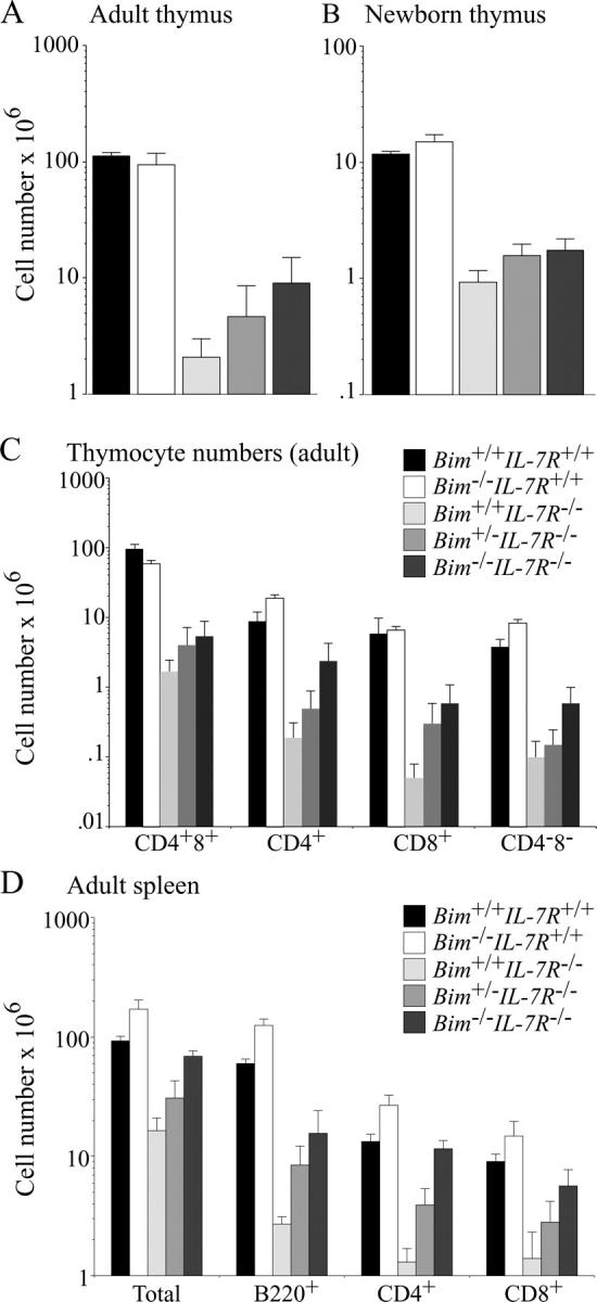
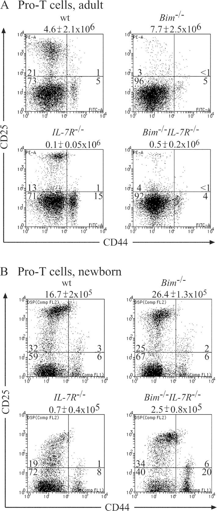
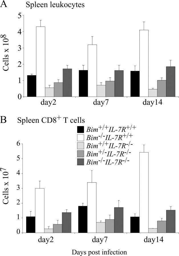
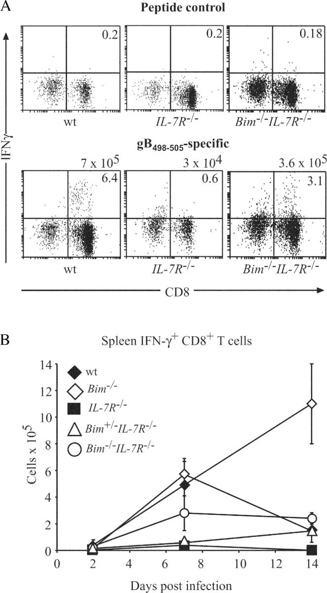
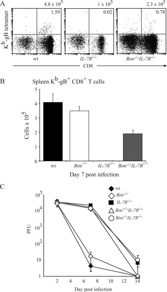
Interleukin (IL)-7 receptor (R) signaling is essential for T and B lymphopoiesis by promoting proliferation, differentiation, and survival of cells. Mice lacking either IL-7 or the IL-7Ralpha chain have abnormally low numbers of immature as well as mature T and B lymphocytes. Transgenic expression of the apoptosis inhibitor Bcl-2 rescues T cell development and function in IL-7Ralpha-deficient mice, indicating that activation of a proapoptotic Bcl-2 family member causes death of immature and mature T cells. BH3-only proteins such as Bim, which are distant proapoptotic members of the Bcl-2 family, are essential initiators of programmed cell death and stress-induced apoptosis. We generated Bim/IL-7Ralpha double deficient mice and found that loss of Bim significantly increased thymocyte numbers, restored near normal numbers of mature T cells in the blood and spleen, and enhanced cytotoxic T cell responses to virus infection in IL-7Ralpha-/- mice. These results indicate that Bim cooperates with other proapoptotic proteins in the death of IL-7-deprived T cell progenitors in vivo, but is the major inducer of this pathway to apoptosis in mature T cells. This indicates that pharmacological inhibition of Bim function might be useful for boosting immune responses in immunodeficient patients.


Figures

Figure 1.
Figure 1.
Loss of Bim increases thymus cellularity and numbers of splenic T and B cells in IL-7Rα−/− mice. 6–10-wk-old (A, C, and D) or newborn (B) WT, Bim −/−, IL-7Rα−/−, Bim +/− IL-7Rα−/−, and Bim −/− IL-7Rα−/− mice were killed to determine total leukocyte numbers in the thymus (A, B, and C) and spleen (D). Cell subset composition was determined by immunofluorescent staining with surface marker–specific antibodies (CD4 and CD8 for T cells and B220, CD19, and sIg for B cells) followed by flow cytometric analysis. Data are presented as means ± SD of at least five mice of each genotype.
Figure 2.
Figure 2.
Effect of loss of Bim on the numbers of pro–T cells (CD348) in the thymus of IL-7Rα−/− mice. 6–10-wk-old (A) or newborn (B) WT, IL-7Rα−/−, Bim −/−, and Bim −/− IL-7Rα−/− mice were killed and thymocytes were stained with Tricolor streptavidin and a cocktail of biotinylated antibodies to exclude immature CD4+8+ thymocytes, mature CD4+8 and CD48+ T cells, B cells, myeloid and erythroid cells, plus a Cy-5–labeled antibody to Thy1 to gate on T lymphoid cells. Pro–T cells were visualized by staining with a PE-labeled anti-CD25 antibody and a FITC-labeled anti-CD44 antibody. Total numbers of thymocytes are listed above the dot plots and the percentages of cells in each of the quadrant are indicated. Data shown are representative of at least five mice of each genotype.
Figure 3.
Figure 3.
Total numbers of leukocytes and CD48+ splenic T cells in HSV-infected WT, Bim −/−, IL-7Rα−/−, Bim +/− IL-7Rα−/−, and Bim −/− IL-7Rα−/− mice. Control WT, Bim −/−, IL-7Rα−/−, Bim +/− IL-7Rα−/−, and Bim −/− IL-7Rα−/− mice were infected with HSV by injection into both hind feet. On days 2, 7, and 14 after infection, total numbers of leukocytes (A) and CD48+ T cells (B) in the spleen were determined by cell counting combined with immunofluorescent staining with surface marker–specific antibodies and FACS analysis. Data shown represent means ± SD of at least three mice of each genotype.
Figure 4.
Figure 4.
Loss of Bim enhances CTL immune responses to HSV in IL-7Rα−/− mice. Control WT, Bim −/−, IL-7Rα−/−, Bim +/− IL-7Rα−/−, and Bim −/− IL-7Rα−/− mice were infected with HSV by injection into both hind feet. (A) On days 2, 7, and 14 after infection, HSV-specific CTLs in the spleen were enumerated by stimulating cells in culture with the gB498–505 peptide (the major epitope from HSV recognized by CTLs in C57BL/6 mice) or as a control with Kb-OVA257–264 peptide SIINFEKL, followed by intracellular staining for IFN-γ. Total numbers of HSV-specific CTLs are listed above the dot plots and the percentages of CD8+ IFN-γ+ T cells are indicated in the top right quadrants. (B) The means ± SD of such analyses using at least three mice of each genotype at each time point are shown.
Figure 5.
Figure 5.
Loss of Bim enhances CTL immune responses to HSV in IL-7Rα−/− mice, but does not accelerate viral clearance. Control WT, Bim −/−, IL-7Rα−/−, Bim +/− IL-7Rα−/−, and Bim −/− IL-7Rα−/− mice were infected with HSV by injection into both hind feet. (A) On day 7 after infection, HSV-specific CTLs in the spleen were enumerated by staining with antibodies to CD8 plus PE-conjugated MHC class I tetrameric complexes incorporating the gB498–505 HSV glycoprotein peptide SSIEFARL, the major epitope from HSV recognized by CD8+ T cells. Total numbers of HSV-specific CTLs are listed above the dot plots and the percentages of CD8+ T cells binding to gB498–505 HSV presented by MHC class I are indicated in the top right quadrants. (B) The means ± SD of such analyses using at least three mice of each genotype are shown. (C) The kinetics of viral clearance in the infected mice as determined by viral plaque assays on extracts from feet are shown. Data shown represent means ± SD of viral PFUs for both feet from at least three mice of each genotype for each time point.

References

    1. Noguchi, M., Y. Nakamura, S.M. Russell, S.F. Ziegler, M. Tsang, X. Cao, and W.J. Leonard. 1993. Interleukin-2 receptor γ chain: a functional component of the interleukin-7 receptor. Science. 262:1877–1880. - PubMed
    1. Morrissey, P.J., P. Conlon, K. Charrier, S. Braddy, A. Alpert, D. Williams, A.E. Namen, and D. Mochizuki. 1991. Administration of IL-7 to normal mice stimulates B-lymphopoiesis and peripheral lymphadenopathy. J. Immunol. 147:561–568. - PubMed
    1. Fisher, A.G., C. Burdet, C. Bunce, M. Merkenschlager, and R. Ceredig. 1995. Lymphoproliferative disorders in IL-7 transgenic mice: expansion of immature B cells which retain macrophage potential. Int. Immunol. 7:415–423. - PubMed
    1. Peschon, J.J., P.J. Morrissey, K.H. Grabstein, F.J. Ramsdell, E. Maraskovsky, B.C. Gliniak, L.S. Park, S.F. Ziegler, D.E. Williams, C.B. Ware, et al. 1994. Early lymphocyte expansion is severely impaired in interleukin 7 receptor–deficient mice. J. Exp. Med. 180:1955–1960. - PMC - PubMed
    1. von Freeden-Jeffry, U., P. Vieira, L.A. Lucian, T. McNeil, S.E.G. Burdach, and R. Murray. 1995. Lymphopenia in interleukin (IL)-7 gene-deleted mice identifies IL-7 as a nonredundant cytokine. J. Exp. Med. 181:1519–1526. - PMC - PubMed

Publication types