. 2004 Oct 25;167(2):377-88.
doi: 10.1083/jcb.200404129.

A transmigratory cup in leukocyte diapedesis both through individual vascular endothelial cells and between them

Affiliations

A transmigratory cup in leukocyte diapedesis both through individual vascular endothelial cells and between them

Christopher V Carman et al. J Cell Biol. .

Abstract

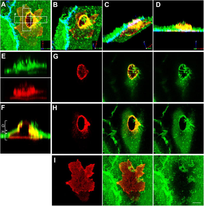
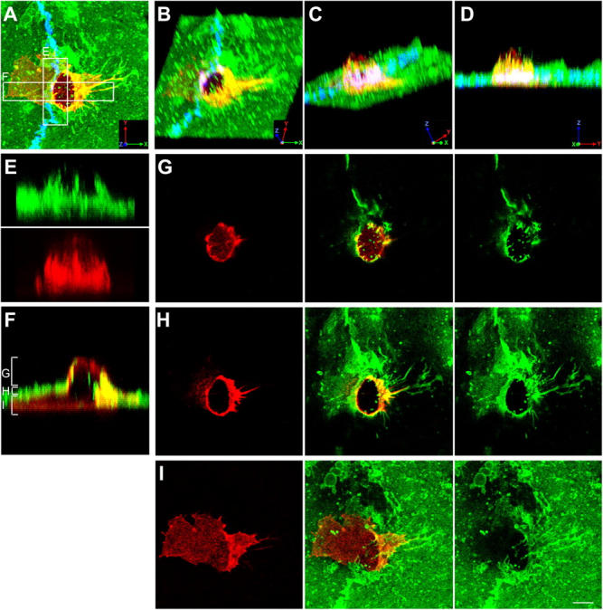
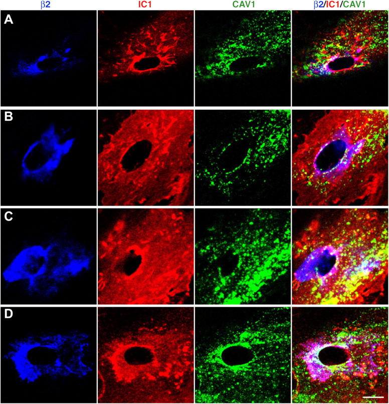
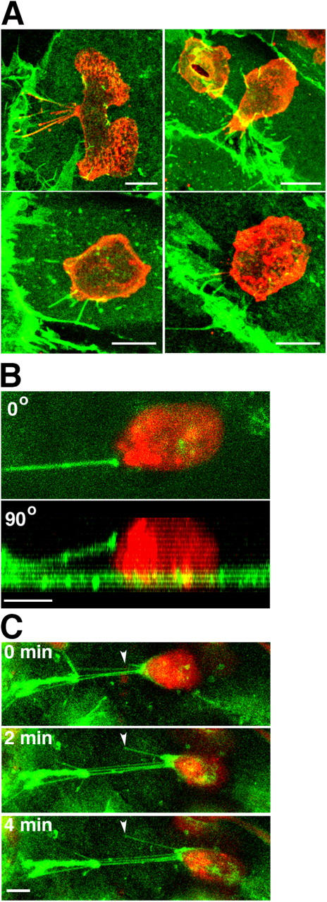
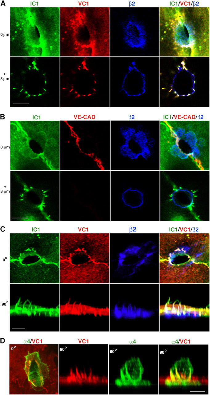
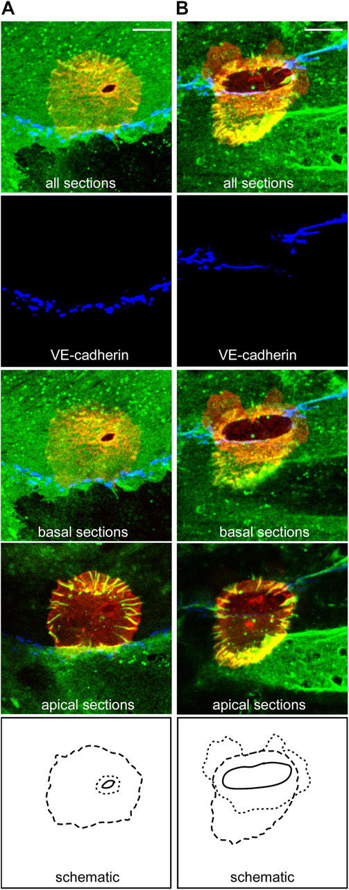
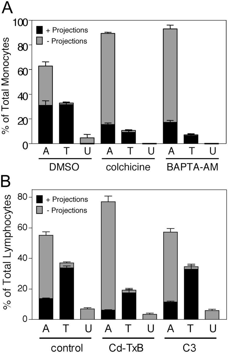
The basic route and mechanisms for leukocyte migration across the endothelium remain poorly defined. We provide definitive evidence for transcellular (i.e., through individual endothelial cells) diapedesis in vitro and demonstrate that virtually all, both para- and transcellular, diapedesis occurs in the context of a novel "cuplike" transmigratory structure. This endothelial structure was comprised of highly intercellular adhesion molecule-1- and vascular cell adhesion molecule-1-enriched vertical microvilli-like projections that surrounded transmigrating leukocytes and drove redistribution of their integrins into linear tracks oriented parallel to the direction of diapedesis. Disruption of projections was highly correlated with inhibition of transmigration. These findings suggest a novel mechanism, the "transmigratory cup", by which the endothelium provides directional guidance to leukocytes for extravasation.


Figures

Figure 1.
Figure 1.
Monocytes migrate across the endothelium via transcellular routes in association with ICAM-1 projections. TNF-α–activated HUVECs were pretreated with MCP-1 and incubated with freshly isolated monocytes for 20 min. Cells were fixed and stained for ICAM-1 (CBRIC1/11-488; green), LFA-1 (CBR-LFA1/7-Cy3; red), and VE-cadherin (55-7H1-Cy5; blue; shown in A–D only) and imaged by confocal microscopy. (A) Top view projection of all z-series sections of a representative monocyte transmigrating via a transcellular route. (B–D) Field in A was rendered as a series of three-dimensional projections, each representing successive rotation about both the x and z axis in 30° intervals for a total of 90° about each axis. (E) Side view projection of cross section E in A depicting ICAM-1 projections (top) and linear LFA-1 clusters (bottom) separately. (F) Side view projection of cross section F in A. Apical (G), middle (H), or basal (I) z-axis sections, as indicated by brackets in F, are projected as top views. Bar, 5 μm.
Figure 2.
Figure 2.
Monocytes migrate across the endothelium via paracellular routes in association with ICAM-1 projections. Samples were prepared as in Fig. 1 with ICAM-1, LFA-1, and VE-cadherin (shown in A only) represented by green, red, and blue fluorescence, respectively. (A) Top view projection of all z-series sections of a representative monocyte transmigrating via a paracellular route. (B–D) Field in A was rendered as a series of three-dimensional projections, each representing successive rotation about both the x and z axis in 30° intervals for a total of 90° about each axis. (E) Side view projection of cross section of E in A depicting ICAM-1 projections (top) and linear LFA-1 clusters (bottom) separately. (F) Side view projection of cross section F in A. Apical (G), middle (H), or basal (I) z-axis sections, as indicated by brackets in F, are projected as top views. Bar, 5 μm.
Figure 3.
Figure 3.
Caveolin-1 is partially associated with the transcellular migration pore. Human lymphocytes were incubated for 10 min with TNF-α–activated, SDF-1–pretreated HUVECs transfected with either caveolin-1-GFP (A and B) or GFP-caveolin-1 (C and D) followed by fixation and staining. β2 (blue), ICAM-1 (IC1; red), and caveolin-1 (cav-1; green) are shown for representative transcellular migration events. Confocal sections that encompass the TEM passage (Fig.1, F and H) are projected as top views. Bar, 5 μm.
Figure 4.
Figure 4.
ICAM-1 projections are highly associated with transmigrating cells. Monocytes (A) and neutrophils and lymphocytes (B) were incubated with TNF-α–activated and either MCP-1–, PAF-, or SDF-1–pretreated HUVEC monolayers, respectively, for the indicated number of minutes (m). Cells were fixed and stained for ICAM-1, LFA-1, and VE-cadherin. In each of three to eight separate experiments, a minimum of 100 leukocytes from randomly selected fields for each time point were carefully analyzed in all apical to basal planes and scored for the presence of ICAM-1–enriched projections of 1 μm in length or greater and as being either apically adherent (A), in the process of TEM (including both para- and transcellular events and including TEM stages 1–3) (T) or under (U) the HUVEC monolayer. Each bar represents the percentage of total cells scored at each indicated time point that were found in each of the three categories (i.e., A, T, or U). The black portion of each bar is the fraction of cells scored positive for associated ICAM-1 projections. The gray portion of each bar is the fraction of the cells that were negative for the presence of ICAM-1 projections. Values represent mean ± SEM (n = 3–8).
Figure 5.
Figure 5.
Asymmetric projections are associated with laterally migrating cells. Monocytes (A) and lymphocytes (B and C) were incubated with TNF-α–activated and either MCP-1– or SDF-1–pretreated HUVEC monolayers, respectively. (A) After 10 min of incubation cells were fixed and stained for ICAM-1 (green) and LFA-1 (red). Four representative top view images depict apically adherent monocytes with asymmetrically distributed ICAM-1 projections. (B) Live-cell imaging of a representative laterally migrating SNARF-labeled lymphocyte (red) on the apical surface of the endothelium prelabeled with an anti–ICAM-1–Alexa 488 Fab fragment (green). Image depicts top (0°; top) and side (90°; bottom) views of a single ICAM-1–bearing cellular projection that is largely suspended above the apical surface of the endothelium and is attached to a lymphocyte migrating laterally to the right. (C) Live-cell time lapse imaging of another cell as in B. ICAM-1 projections emanating from the apical surface of the endothelium (arrowheads) attached to the trailing edge of a lymphocyte become elongated as the lymphocyte advances. Panels depict top view projections of confocal sections at 0, 2, and 4 min as indicated. Bars, 5 μm.
Figure 6.
Figure 6.
ICAM-1 and VCAM-1, but not VE-cadherin, are highly enriched in projections surrounding sites of transmigration. (A) Top view of 0.2-μm-thick confocal sections of a monocyte in TEM-1 at 0 μm (top) and 3 μm (bottom) above the apical surface of the endothelium. ICAM-1 (IC1, green), VCAM-1 (VC1, red), and β2 integrin (β2, blue) are shown either separately (left three) or merged (right). (B) Top view of confocal sections of a monocyte in TEM-1 at 0 μm (top) and 3 μm (bottom) above the apical endothelial surface. ICAM-1 (IC1, green), VE-cadherin (VE-CAD, red), and β2 integrin (β2, blue) are shown either separately (left three) or merged (right). Note that VE-cadherin staining at 0 μm is continuous at the TEM passage in contrast to Fig. S4 C. (C) Serial confocal sections of a monocyte in TEM-2 projected as top (0o, top) or side (90°, bottom) views. ICAM-1 (IC1, green), VCAM-1 (VC1, red), and β2 integrins (β2, blue) are shown either separately (left three) or merged (right). Three-dimensional rotation of these projections is shown in Video 1. (D) Serial confocal sections of a lymphocyte in TEM-2 projected as top (0o, left) or side (90°, right three) views. α4 integrin (α4, green) and VCAM-1 (VC1, red) are shown either separately (middle) or merged (left and right). Bars, 5 μm.
Figure 7.
Figure 7.
At early stages of TEM ICAM-1 projections encircle the endothelial migration passage. Monocytes were incubated with TNF-α–activated, MCP-1–pretreated HUVECs for 10–20 min and then fixed and stained for ICAM-1 (IC1/11-488, green), LFA-1 (CBR-LFA1/7-Cy3, red), and VE-cadherin (55-7H1-Cy5, blue) and imaged by confocal microscopy. (A) All sections, the basal sections or the apical sections of a representative monocyte at an early stage of transcellular TEM-1. (B) A representative monocyte at a late stage of paracellular TEM-1. Dotted, dashed, and solid lines shown in the bottom panels indicate the edges of the sub-endothelial leukocyte membrane, the perimeter of the ICAM-1 projections and the TEM passage, respectively. Note that the TEM passage resides within the perimeter established by the ICAM-1 projections. Bars, 5 μm.
Figure 8.
Figure 8.
Inhibition of ICAM-1 projections by BAPTA-AM, colchicine, or toxin-B is highly correlated with reduced TEM. (A) TNF-α–activated, MCP-1–pretreated HUVECs were pretreated with vehicle (DMSO, 60 min), 10 μm colchicine (20 min), or 20 μm BAPTA-AM (60 min) washed and incubated with monocytes for 10 min at 37°C. Cells were fixed, stained for ICAM-1 (IC1/11-488), and LFA-1 (CBR-LFA1/7-Cy3) and then analyzed by confocal microscopy. In each of three to five separate experiments at least 100 monocytes in randomly selected fields were carefully analyzed in all apical to basal planes and scored for the presence of significant ICAM-1–enriched projections of 1 μm in length or greater and as being either apically adherent (A), in the process of TEM (including both para- and transcellular events and TEM 1–3) (T) or under (U) the HUVEC monolayer. Each bar represents the percentage of total cells scored for each experimental condition (DMSO, colchicine, or BAPTA-AM) in each of the three categories. The black portion of each bar is the fraction of cells positive for associated ICAM-1 projections. The gray portion of each bar is the fraction of the cells that were negative for the presence of ICAM-1 projections. (B) TNF-α–activated, SDF-1–pretreated HUVECs were pretreated with vehicle (PBS, 60 min, control), 100 ng/ml toxin-B (60 min) or 50 μg/ml C3 transferase (16 h) washed and then incubated with human lymphocytes for 10 min at 37°C. Samples were fixed, stained, and analyzed as in A. Values represent mean ± SEM (n = 3–5).

References

    1. Adamson, P., S. Etienne, P.O. Couraud, V. Calder, and J. Greenwood. 1999. Lymphocyte migration through brain endothelial cell monolayers involves signaling through endothelial ICAM-1 via a rho-dependent pathway. J. Immunol. 162:2964–2973. - PubMed
    1. Aurrand-Lions, M., C. Johnson-Leger, and B.A. Imhof. 2002. The last molecular fortress in leukocyte trans-endothelial migration. Nat. Immunol. 3:116–118. - PubMed
    1. Baekkevold, E.S., T. Yamanaka, R.T. Palframan, H.S. Carlsen, F.P. Reinholt, U.H. von Andrian, P. Brandtzaeg, and G. Haraldsen. 2001. The CCR7 ligand elc (CCL19) is transcytosed in high endothelial venules and mediates T cell recruitment. J. Exp. Med. 193:1105–1112. - PMC - PubMed
    1. Barreiro, O., M. Yanez-Mo, J.M. Serrador, M.C. Montoya, M. Vicente-Manzanares, R. Tejedor, H. Furthmayr, and F. Sanchez-Madrid. 2002. Dynamic interaction of VCAM-1 and ICAM-1 with moesin and ezrin in a novel endothelial docking structure for adherent leukocytes. J. Cell Biol. 157:1233–1245. - PMC - PubMed
    1. Bianchi, E., J.R. Bender, F. Blasi, and R. Pardi. 1997. Through and beyond the wall: late steps in leukocyte transendothelial migration. Immunol. Today. 18:586–591. - PubMed

Publication types

MeSH terms