Case Reports
. 2004 Oct 26;63(8):1376-84.
doi: 10.1212/01.wnl.0000139809.16817.dd.

Clinical and neuropathologic variation in neuronal intermediate filament inclusion disease

Affiliations
Case Reports

Clinical and neuropathologic variation in neuronal intermediate filament inclusion disease

N J Cairns et al. Neurology. .

Abstract

Background: Recently described neuronal intermediate filament inclusion disease (NIFID) shows considerable clinical heterogeneity.

Objective: To assess the spectrum of the clinical and neuropathological features in 10 NIFID cases.

Methods: Retrospective chart and comprehensive neuropathological review of these NIFID cases was conducted.

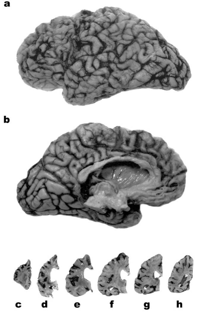
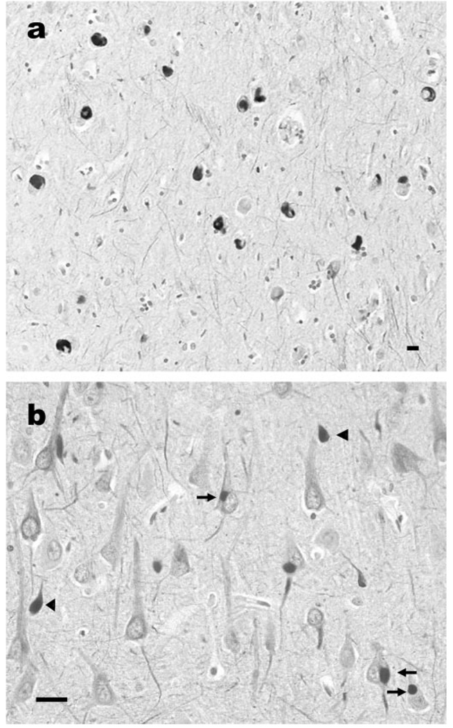
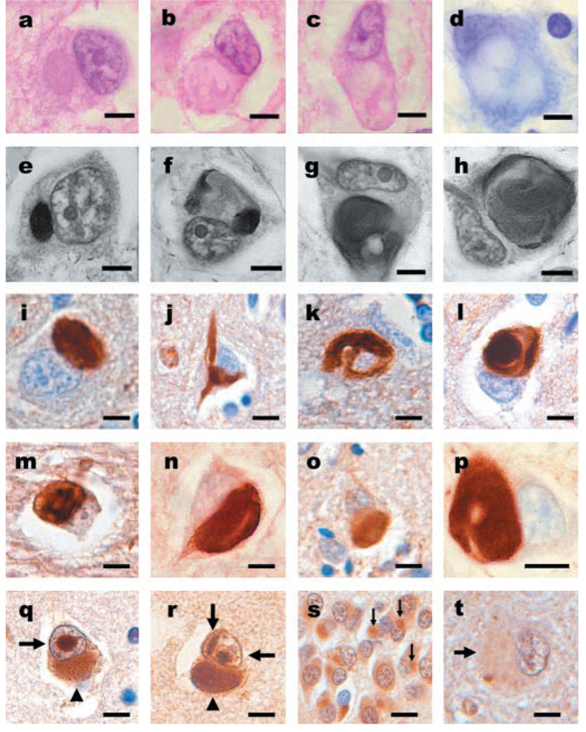
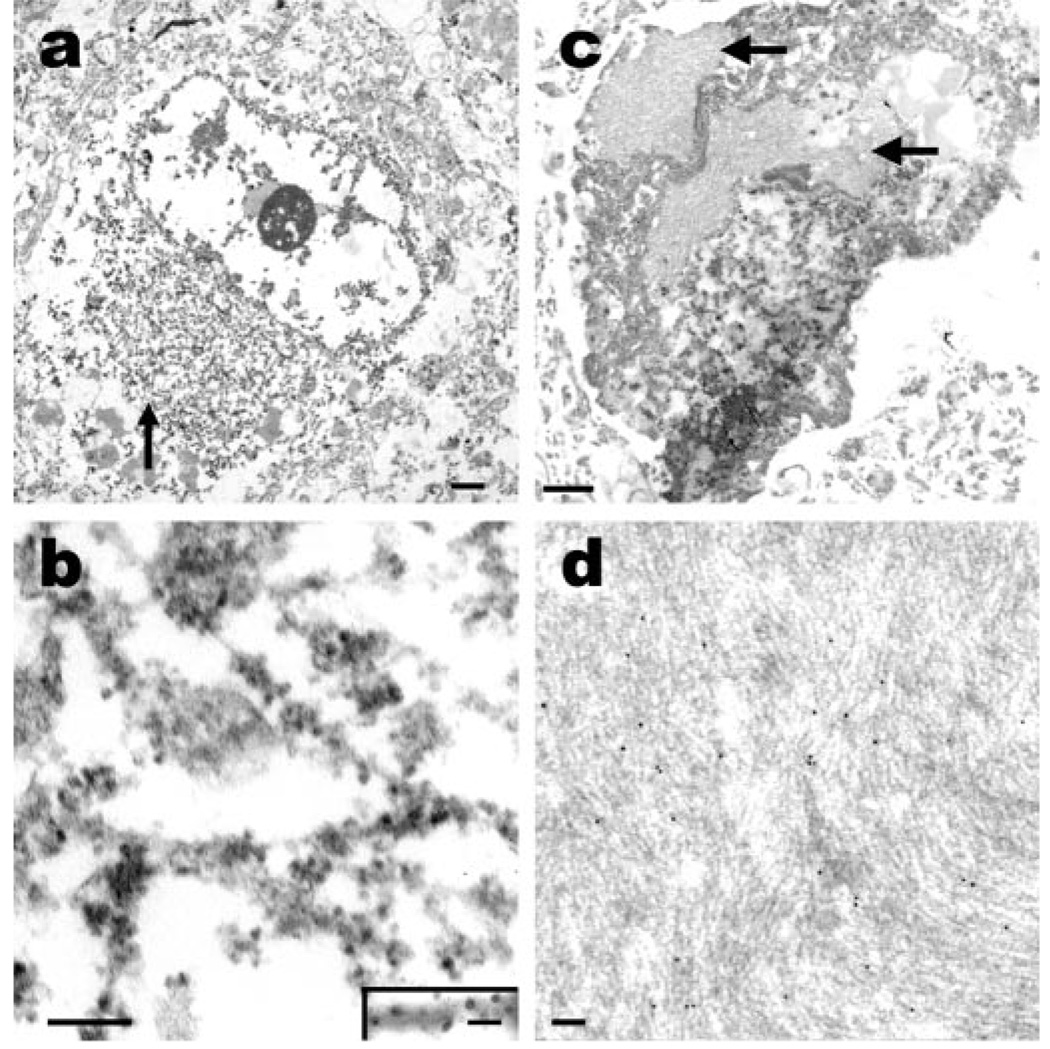
Results: The mean age at onset was 40.8 (range 23 to 56) years, mean disease duration was 4.5 (range 2.7 to 13) years, and mean age at death was 45.3 (range 28 to 61) years. The most common presenting symptoms were behavioral and personality changes in 7 of 10 cases and, less often, memory loss, cognitive impairment, language deficits, and motor weakness. Extrapyramidal features were present in 8 of 10 patients. Language impairment, perseveration, executive dysfunction, hyperreflexia, and primitive reflexes were frequent signs, whereas a minority had buccofacial apraxia, supranuclear ophthalmoplegia, upper motor neuron disease (MND), and limb dystonia. Frontotemporal and caudate atrophy were common. Histologic changes were extensive in many cortical areas, deep gray matter, cerebellum, and spinal cord. The hallmark lesions of NIFID were unique neuronal IF inclusions detected most robustly by antibodies to neurofilament triplet proteins and alpha-internexin.

Conclusion: NIFID is a neuropathologically distinct, clinically heterogeneous variant of frontotemporal dementia (FTD) that may include parkinsonism or MND. Neuronal IF inclusions are the neuropathological signatures of NIFID that distinguish it from all other FTD variants including FTD with MND and FTD tauopathies.


Figures

Figure 1
Figure 1
The lateral (a) and medial (b) aspects of the left hemibrain of 27-year-old woman with neuronal intermediate filament inclusion disease. There is pronounced atrophy of the frontal and anterior temporal lobes. Coronal slices of the left hemisphere (c through h) reveal enlargement of the lateral ventricle, and marked atrophy of the striatum (d). The Sylvian fissure (e) is widened and the amygdala (e) and hippocampus (f, g) are atrophied. The parietal (a, b, h) and occipital (a, b) lobes are relatively well preserved.
Figure 2
Figure 2
Neuronal intermediate filament inclusions in the temporal lobe of a case of neuronal intermediate filament inclusion disease. There are pleomorphic neuronal inclusions in the subiculum (a) and in the perikarya of pyramidal neurons (b, arrows) and in swollen axons and spheroids (b, arrowheads) of hippocampal subfield CA1. α-Internexin immunohistochemistry. Scale bar = 10 µm.
Figure 3
Figure 3
Neuronal inclusions in neuronal intermediate filament inclusion disease (NIFID) are pleomorphic and contain all class IV IF proteins. (a) An eosinophilic, cytoplasmic inclusion in the frontal lobe. (b, c) Neurons with globular or elongated inclusions with hyaline compartments of variable eosinophilia. (a through c) Hematoxylin and eosin. (d) Only the central core of a hyaline inclusion is visible with cresyl violet stain. (e through h) Inclusions are variably argyrophilic and pleomorphic: some are spheroidal (a), others have stained and unstained compartments (f), and others have serpiginous and complex forms (g, h). Modified Bielschowsky silver impregnation. (i) Pick body-like inclusions are the most common morphological type. Inclusions contain all neuronal IF proteins. (j) A flame-shaped inclusion. (k) A filamentous serpiginous inclusion. (l) A globose inclusion. (i through l) α-Internexin immunohistochemistry. Epitopes of phosphorylation-dependent NF-H (RMO 24) (m), non-phosphorylation-dependent NF-H (RMd 09) (n), phosphorylation-independent NF (NF-M; RMO 189) (o), phosphorylation-independent NF (NF-L; NR4) (p), are present in the inclusions. (m through p) Neurofilament immunohistochemistry. Neuronal inclusions in NIFID are variably ubiquitinated. (q, r) Neuronal cytoplasmic (arrowhead) and intranuclear inclusions (arrows) are present in the same neurons. The intranuclear inclusions appear as round or elongated structures either as a single inclusion (q) or as multiple forms (r). (s) Inclusions in the neurons of the dentate gyrus are ubiquitinated (arrows). (t) In a 61-year-old case of NIFID, a hyaline neuronal fibrillary inclusion is weakly stained. (q through t) Ubiquitin immunohistochemistry. (a through h) Scale bars = 5 µm; (i through t) scale bars = 10 µm.
Figure 4
Figure 4
Fine structure of inclusions in NIFID. (a) An electron micrograph of a neuronal cytoplasmic inclusion (arrow) in the frontal lobe. The inclusion is not membrane bound and contains granular filamentous material with a diameter of 10 to 25 nm (b). Immunoelectron microscopy (b, inset) reveals that the filaments of the inclusion contain epitopes of heavy-subunit neurofilament (RMO 24, immunoperoxidase-labeling). Hyaline bodies in NIFID contain dense fibrillar arrays. (c) A pyknotic neuron containing hyaline inclusions of closely packed arrays of filaments (arrows). (d) Immunoelectron microscopy of hyaline bodies reveals that the filaments contain epitopes of neurofilament proteins (SMI 312 labeled with 10-nm gold particles). (a, c) Scale bar = 1 µm; (b, d) scale bar = 100 nm and (b, inset) scale bar = 20 nm.

Comment in

References

    1. Cairns NJ, Perry RH, Jaros E, et al. Patients with a novel neurofilamantopathy: dementia with neurofilament inclusions. Neurosci Lett. 2003;341:177–180. - PubMed
    1. Cairns NJ, Zhukareva V, Uryu K, et al. α-Internexin is present in the pathological inclusions of neuronal intermediate filament inclusion disease. Am J Pathol. 2004;164:2153–2161. - PMC - PubMed
    1. Cairns NJ, Uryu K, Bigio E, et al. α-Internexin aggregates are abundant in neuronal intermediate filament inclusion disease (NIFID) but rare in other neruodegenerative diseases. Acta Neuropathol. 2004;108:213–233. - PMC - PubMed
    1. Bigio EH, Lipton AM, White CL, III, et al. Frontotemporal dementia and motor neurone degeneration with neurofilament inclusion bodies: additional evidence for overlap between FTD and ALS. Neuropathol Appl Neurobiol. 2003;29:239–253. - PubMed
    1. Duyckaerts C, Mokhtari K, Fontaine B, et al. Maladie de Pick généralisée: une démence mal nommée caractérisée par des inclusions neurofilamentaires. Rev Neurol (Paris) 2003;159:219–219.

Publication types

MeSH terms