Comparative Study
. 2004 Oct 26;63(8):1393-8.
doi: 10.1212/01.wnl.0000141849.21256.ac.

1H MR spectroscopy in common dementias

Affiliations
Comparative Study

1H MR spectroscopy in common dementias

K Kantarci et al. Neurology. .

Abstract

Objective: To determine the 1H MR spectroscopic (MRS) findings and intergroup differences among common dementias: Alzheimer disease (AD), vascular dementia (VaD), dementia with Lewy bodies (DLB), and frontotemporal lobar degeneration (FTLD).

Methods: The authors consecutively recruited 206 normal elderly subjects and 121 patients with AD, 41 with FTLD, 20 with DLB, and 8 with VaD. The 1H MRS metabolite ratio changes in common dementias were evaluated with respect to normal and also differences among the common dementias.

Results: N-acetylaspartate (NAA)/creatine (Cr) was lower than normal in patients with AD, FTLD, and VaD. Myo-inositol (mI)/Cr was higher than normal in patients with AD and FTLD. Choline (Cho)/Cr was higher than normal in patients with AD, FTLD, and DLB. There were no metabolite differences between patients with AD and FTLD or between patients with DLB and VaD. NAA/Cr was lower in patients with AD and FTLD than DLB. MI/Cr was higher in patients with AD and FTLD than VaD. MI/Cr was also higher in patients with FTLD than DLB.

Conclusions: NAA/Cr levels are decreased in dementias that are characterized by neuron loss, such as AD, FTLD, and VaD. MI/Cr levels are elevated in dementias that are pathologically characterized by gliosis, such as AD and FTLD. Cho/Cr levels are elevated in dementias that are characterized by a profound cholinergic deficit, such as AD and DLB.


Figures

Figure 1
Figure 1
Placement of the 1H MRS voxel on a mid-sagittal T1 weighted image. The 8 cm3 (2×2×2cm) voxel is covering right and left hemispheric posterior cingulate gyri and inferior precunei.
Figure 2
Figure 2
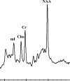
Examples of proton spectra obtained from the posterior cingulate voxel with an echo time of 30 ms. in a normal subject (a), a patient with AD (b), FTLD (c), DLB (d), and VaD (e). The NAA /Cr ratio is lower in patients with AD, FTLD and VaD than both the DLB and the normal subjects. The Cho /Cr ratio is higher in patients with AD, FTLD and DLB than the normal subject. The mI /Cr ratio is higher in the patients with AD and FTLD than both the VaD and the normal subjects. All spectra are scaled to the height of the reference peak Cr, which is held constant across Figures 2a-2e.
Figure 2
Figure 2
Examples of proton spectra obtained from the posterior cingulate voxel with an echo time of 30 ms. in a normal subject (a), a patient with AD (b), FTLD (c), DLB (d), and VaD (e). The NAA /Cr ratio is lower in patients with AD, FTLD and VaD than both the DLB and the normal subjects. The Cho /Cr ratio is higher in patients with AD, FTLD and DLB than the normal subject. The mI /Cr ratio is higher in the patients with AD and FTLD than both the VaD and the normal subjects. All spectra are scaled to the height of the reference peak Cr, which is held constant across Figures 2a-2e.

Comment in

References

    1. Holmes C, Cairns N, Lantos P, Mann A. Validity of current clinical criteria for Alzheimer's disease, vascular dementia and dementia with Lewy bodies. British Journal of Psychiatry. 1999;174:45–50. - PubMed
    1. Lim A, Tsuang D, Kukull W, et al. Clinico-neuropathological correlation of Alzheimer's disease in a community-based case series. Journal of American Geriatrics Society. 1999;47:564–569. - PubMed
    1. Klunk WE, Panchalingam K, Moosy J, Mc Clure RJ, Pettegrew JW. N-acetyl-L-aspartate and other amino acid metabolites in Alzheimer's disease brain: a preliminary proton nuclear magnetic resonance study. Neurology. 1992;42:1578–1585. - PubMed
    1. Miller BL, Moats RA, Shonk T, Earnst T, Wooley S, Ross BD. Alzheimer disease: Depicition of increased cerebral myo-inositol with proton MR spectroscopy. Radiology. 1993;187:433–437. - PubMed
    1. Schuff N, Amend DL, Meyerhoff DJ, et al. Alzheimer disease: Quantitative H-1 MR Spectroscopic imaging of frontoparietal brain. Radiology. 1998;207:91–102. - PMC - PubMed

Publication types

MeSH terms