. 2004 Nov 2;101(44):15656-60.
doi: 10.1073/pnas.0405429101. Epub 2004 Oct 25.

Nodal protein processing and fibroblast growth factor 4 synergize to maintain a trophoblast stem cell microenvironment

Affiliations

Nodal protein processing and fibroblast growth factor 4 synergize to maintain a trophoblast stem cell microenvironment

Marcela Guzman-Ayala et al. Proc Natl Acad Sci U S A. .

Abstract

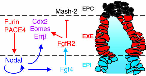
Before implantation in the uterus, mammalian embryos set aside trophoblast stem cells that are maintained in the extraembryonic ectoderm (ExE) during gastrulation to generate the fetal portion of the placenta. Their proliferation depends on diffusible signals from neighboring cells in the epiblast, including fibroblast growth factor 4 (Fgf4). Here, we show that Fgf4 expression is induced by the transforming growth factor beta-related protein Nodal. Together with Fgf4, Nodal also acts directly on neighboring ExE to sustain a microenvironment that inhibits precocious differentiation of trophoblast stem cells. Because the ExE itself produces the proteases Furin and PACE4 to activate Nodal, it represents the first example, to our knowledge, of a stem cell compartment that actively maintains its own microenvironment.


Figures

Fig. 1.
Fig. 1.
Genetic inactivation of Furin and PACE4 or Nodal accelerates the differentiation of TSCs. (A) Sagittal sections through the extraembryonic region on E7.5 shows an expansion of EPC at the expense of the chorion (ch) in Furin/PACE4-deficient DKO embryos (Right), compared with compound heterozygotes (Left). The mutant embryo is also shown at low magnification (the left portion at Right) to indicate the junction between the epiblast and the enlarged EPC (stippled lines). The high magnification (right portion at Right) corresponds to the boxed area. (B) On day E7.5, expression of the EPC marker Mash2 is expanded toward the epiblast boundary (stippled lines) in both DKO (second and third columns) and Nodal mutants (fourth column), whereas progenitor cells marked by Errβ and Cdx2 mRNA in the ExE and chorion are reduced or absent. (Bottom) One day earlier (E6.5), Fgf4 expression in the epiblast was down-regulated compared with control litter mates (first column).
Fig. 2.
Fig. 2.
Processed Nodal stimulates autoinduction and expression of Fgf4. (A) Intact embryos dissected at E5.75 (Left) were cut twice (stippled lines) before or after enzymatic digestion of the overlying VE layer to culture isolated epiblast (Epi) and ExE explants. (B) Compared with cultured whole embryos (WE), epiblast explants deprived of ExE and VE show reduced expression levels of a NodallacZ reporter allele. Background staining in Ndl+/+ explants lacking the lacZ allele is negligible. Ndl, Nodal. (C) The cleaved, mature form of Nodal (filled rectangle) in conditioned medium of transfected 293T cells (Left, lane 2) amplifies expression of the NodallacZ reporter allele (Center), whereas an SPC-resistant mutant precursor precursor (lane 1) has no effect. (D) Recombinant Nodal can also rescue expression of Fgf4, whereas conditioned medium of untransfected, parental 293T cells (mock) was inactive.
Fig. 3.
Fig. 3.
The combined action of epiblast-derived Nodal and FGF4 inhibits precocious differentiation of the ExE. (A) In ExE explants, removal of the epiblast and EPC results in ectopic expression of Mash2 at the expense of Errβ, Cdx2, and Eomesodermin. For clarity, the boundaries of the ExE in whole embryos (WE) are outlined. (B) FGF4 prevents ectopic expression of Mash2, and, if added together with Nodal, induces expression of Errβ, Eomes, and Cdx2 in ExE explants. Note that Nodal alone has no significant effect on Mash2. Furthermore, in negative control experiments, none of the markers examined is influenced by conditioned medium devoid of Nodal (mock).
Fig. 4.
Fig. 4.
Summary of the reciprocal inductive interactions with the epiblast that are required to prevent precocious differentiation of the ExE into EPC tissue. The cartoon depicts a sagittal section (0.8 μm) of a fixed embryo on E6.5 that served as a template (for clarity, the overlying VE layer is omitted). In this model, epiblast (EPI)-derived Nodal and Fgf4 (blue) establish a microenvironment that sustains TSCs in the adjacent ExE (red). To date, no firm evidence exists for any of the available markers that TSCs represent only a subset of ExE cells. However, the model does not exclude the possibility that some TSCs may become biased toward differentiation already within the ExE, e.g., if their apical surface exposed to amniotic fluid is below a critical size.

References

    1. Tanaka, S., Kunath, T., Hadjantonakis, A. K., Nagy, A. & Rossant, J. (1998) Science 282, 2072-2075. - PubMed
    1. Uy, G. D., Downs, K. M. & Gardner, R. L. (2002) Development (Cambridge, U.K.) 129, 3913-3924. - PubMed
    1. Rossant, J., Gardner, R. L. & Alexandre, H. L. (1978) J. Embryol. Exp. Morphol. 48, 239-247. - PubMed
    1. Johnson, M. H. & Rossant, J. (1981) J. Embryol. Exp. Morphol. 61, 103-116. - PubMed
    1. Carney, E. W., Prideaux, V., Lye, S. J. & Rossant, J. (1993) Mol. Reprod. Dev. 34, 357-368. - PubMed

Publication types

MeSH terms